Wavelength Pearls
From the scanning room.
Free, five-minute point-of-care ultrasound pearls from the Wavelength faculty. One technique, one view or one pitfall at a time, each with a short test.
140 pearls in 8 topics. Each teaches one technique and ends with a short test.
Open the question bankFundamentals
Set up the machine and the image before you look for pathology.
Core skills
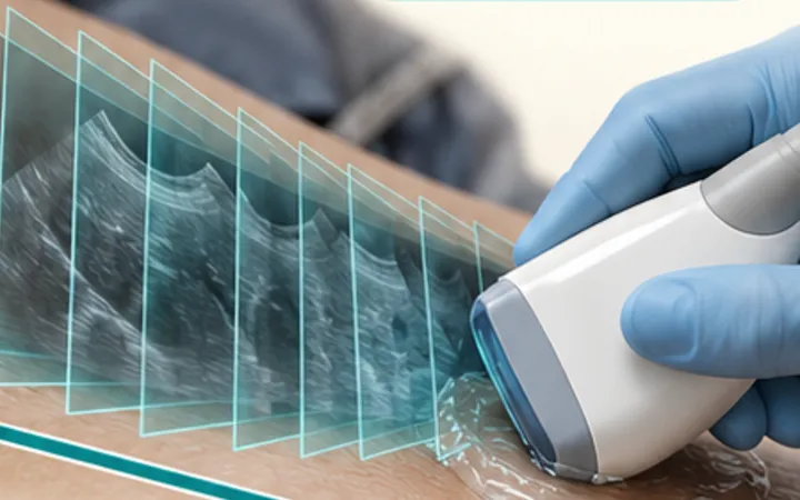
Sweep, fan, slide: do not trust the still image
One frame shows one thin slice. Pathology often sits a few millimetres away. Move the probe through the whole target, by sweeping across it, fanning through it and sliding along it, before you call anything normal.
Core skills
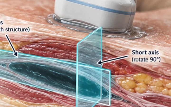
Two planes: long and short or it did not happen
A finding in one plane can be an artefact or a trick of the angle. Rotate the probe 90 degrees. A real vessel, collection or lesion makes anatomical sense in both long and short axis; an artefact usually does not.
Core skills
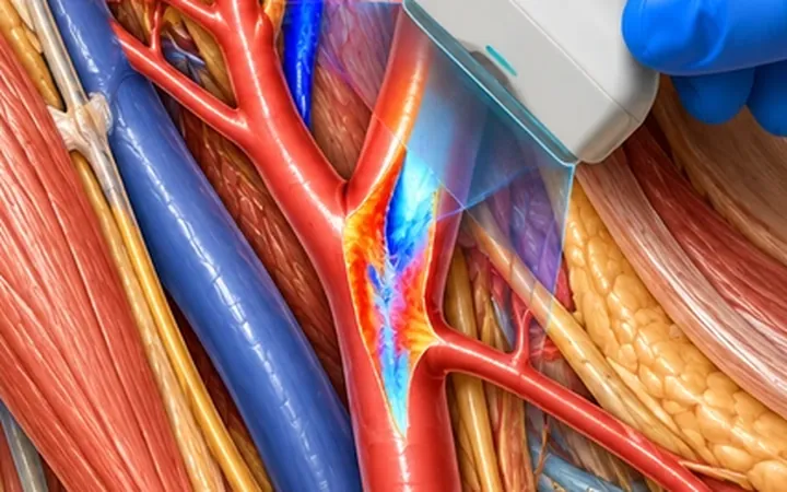
Colour aliasing: change the scale first
Red and blue mixing inside a vessel may be fast flow, or simply a colour scale set too low. Raise the scale, adjust the gain and the angle, then see whether a focal jet is still there.
Core skills
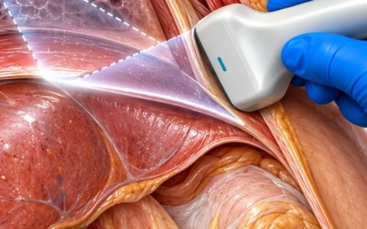
Mirror artefact: read the diaphragm
A copy of the liver or spleen above the diaphragm is a mirror artefact, and it is normal: it means air-filled lung sits above. When the copy disappears and the spine shows above the diaphragm, look for fluid.
Core skills
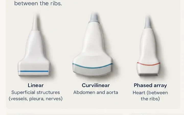
Five checks before every scan
Probe, preset, depth, gain and focus. Thirty seconds of set-up decides whether the image answers your question.
Heart and vessels
Cardiac views, the venous system, the aorta and the leg veins.
Aorta
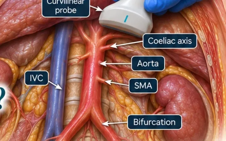
Aorta: when is the scan complete?
An aortic scan is complete only when you follow the aorta from below the diaphragm to the bifurcation. Anything less is an incomplete scan, not a normal one.
Cardiac
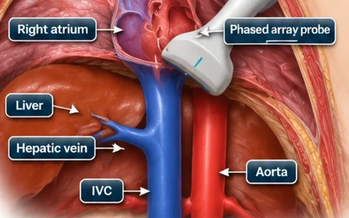
IVC: follow it into the right atrium
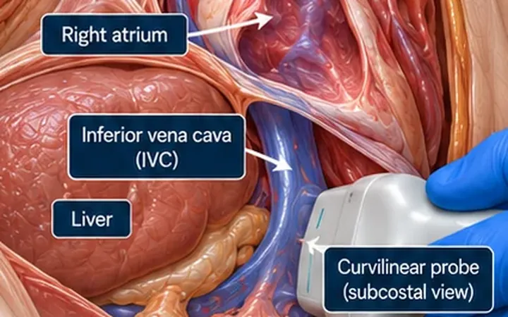
A tube behind the liver is not always the IVC. Start in the subcostal view, keep the right atrium on screen and rotate, and you know you have the right vessel before you measure it.
Cardiac
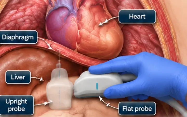
Subcostal view: flatten the probe
Hold the probe upright under the ribs and the beam goes to the spine. Put your hand on top and lay the probe almost flat, and the beam slides under the ribs to the heart.
Vascular
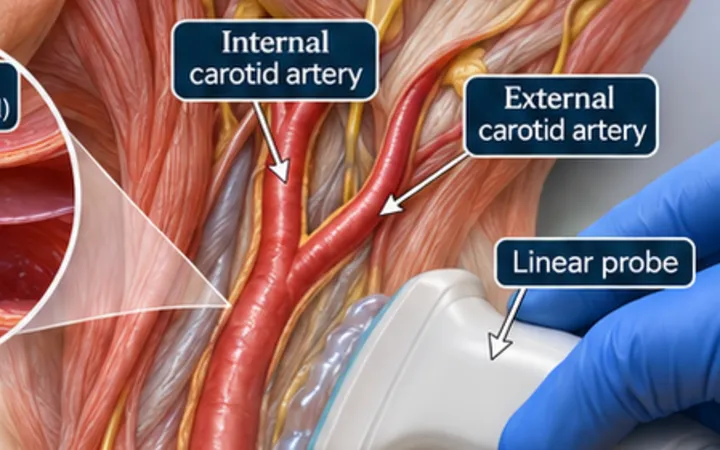
Carotid dissection: look for the intimal flap
Neck pain or headache with a Horner syndrome or focal neurology in a younger patient can be a carotid dissection. A linear probe can show an intimal flap, a double lumen or a crescent of mural haematoma narrowing the lumen. Most dissections start above the reach of the probe, so a normal scan never replaces CT or MR angiography and the stroke pathway.
Cardiac
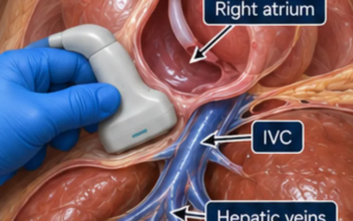
IVC clot: not every big IVC is congestion
A wide IVC is usually read as high right-sided pressure. Before you do, look inside it. Clot or tumour within the IVC also makes it wide and still. If you find it, define how far it extends, especially into the right atrium.
Cardiac
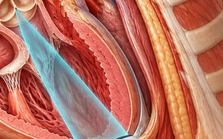
LVOT VTI: trend the same window
LVOT VTI is most useful as a repeatable trend. Keep the view, the sample position, the tracing method and the rhythm the same before you decide that stroke volume has changed.
Cardiac
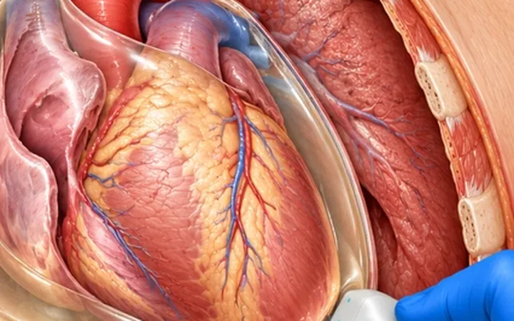
Tamponade: time the collapse
A chamber wall moving inward is not enough. Right atrial collapse matters when it lasts through systole, and right ventricular collapse when it happens in early diastole. Name the phase before you name the sign.
Cardiac
IVC: a big vein is not a fluid order
IVC size and collapse estimate right atrial pressure. They do not tell you whether fluid will help. Read the IVC with the heart, the lungs and the patient.
Cardiac
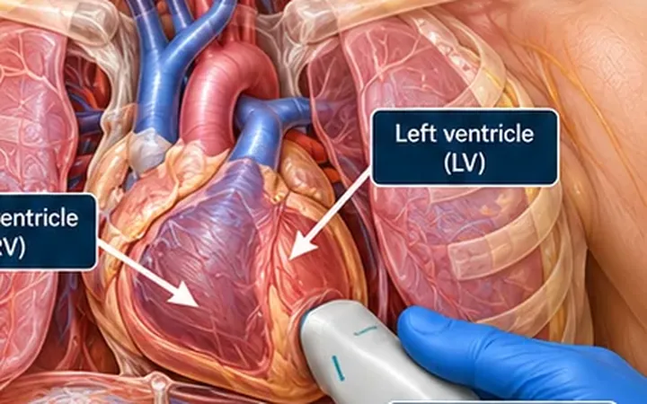
RV strain: three clues beat one ratio
A big right ventricle is a clue. A big right ventricle with a flattened septum, a plethoric IVC and a clot in the leg is a pattern. Build the pattern before you act on it.
Vascular
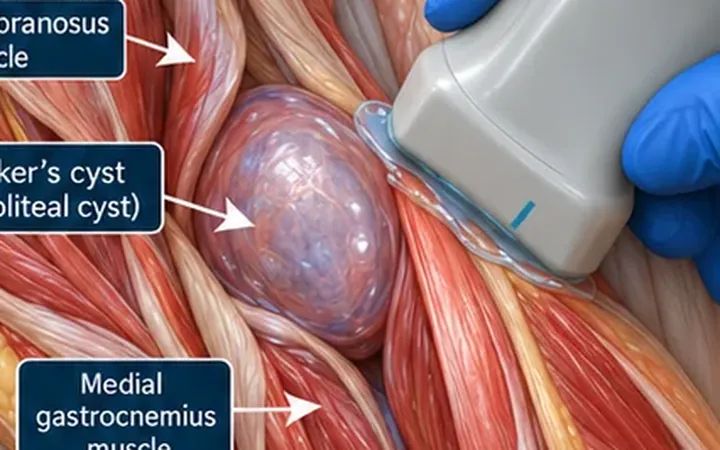
Calf swelling: look behind the knee
A ruptured Baker's cyst mimics a DVT. Find the cyst between semimembranosus and the medial head of gastrocnemius, follow any leaked fluid down the calf, then compress the veins anyway.
Vascular
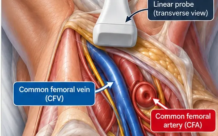
DVT: compress, don't admire
Fresh clot is often black on screen, so a vein full of thrombus looks like a normal vein. The test is whether the walls meet when you press, in transverse, at every site.
Cardiac
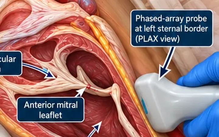
EPSS: when the mitral valve misses the septum
In a strong heart, the anterior mitral leaflet swings open almost to the septum. In a weak heart, it falls short. Measure the gap in the parasternal long-axis view for a quick, objective clue to left ventricular function.
Cardiac
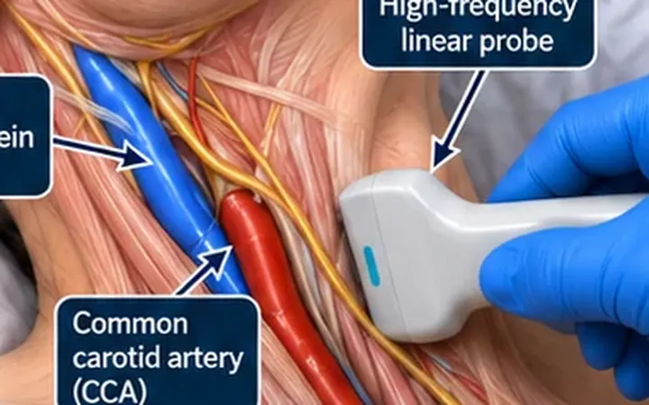
Ultrasound JVP: find the collapse point
The jugular venous pressure is hard to see in a large neck, a busy room or a breathless patient. Follow the internal jugular vein up from the clavicle until it collapses. That point is the top of the venous column.
Cardiac
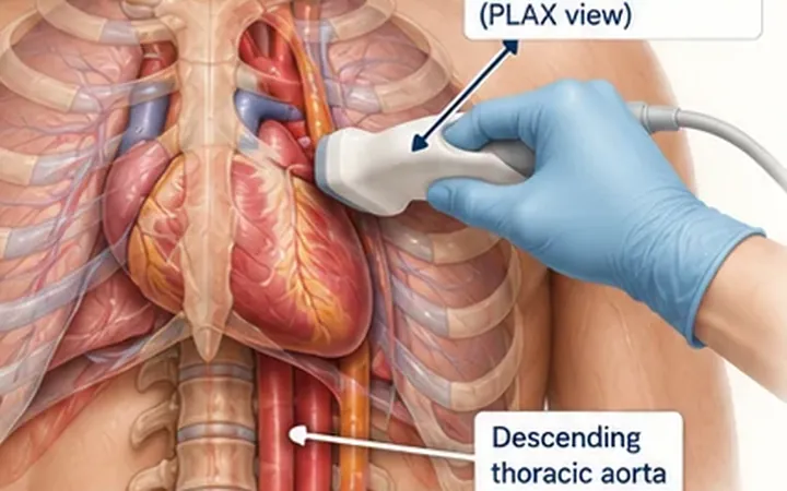
Pericardial or pleural? Find the descending aorta
Black fluid behind the heart in the parasternal long-axis view is either pericardial or pleural. The descending thoracic aorta tells you which. Pericardial fluid runs in front of it. Pleural fluid stays behind it.
Cardiac
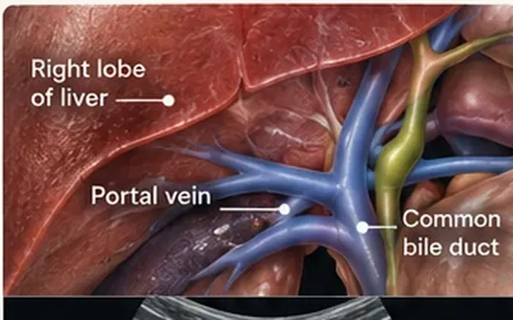
Portal vein pulsatility: a congestion clue
Portal vein flow is normally steady. When pressure backs up from the right heart, it starts to pulse. Read it alongside the IVC, hepatic veins and kidneys, not on its own.
Vascular
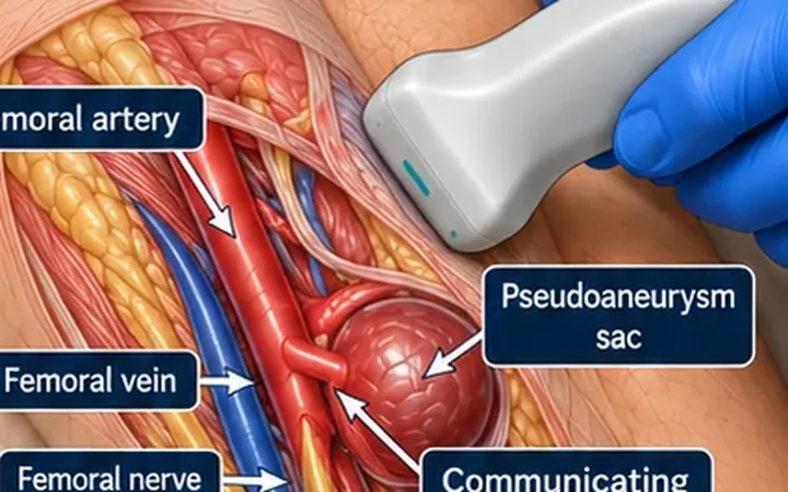
Pseudoaneurysm: find the neck
A pulsatile lump after a femoral puncture is a haematoma, an abscess or a pseudoaneurysm. The swirling sac catches your eye, but the neck makes the diagnosis. Find the channel to the artery and sample it.
Lung and airway
Pneumothorax, consolidation, effusion, the diaphragm and tube position.
Lung
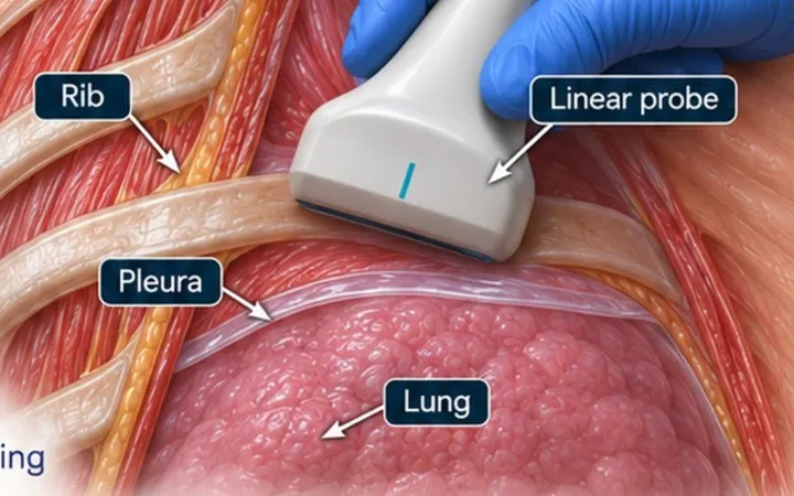
Lung: find the bat before you judge sliding
Lie the probe along a rib and you see one bright line with black below it. Turn it across two ribs first, and the pleural line sits between the rib shadows where you can trust it.
Airway
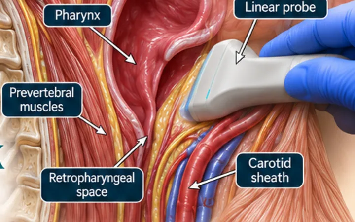
Retropharyngeal abscess: look behind the pharynx
Fever, neck pain, a stiff neck that will not extend, drooling or stridor in a young child can be a retropharyngeal abscess. In a calm patient, a linear probe on the side of the neck can sometimes show a collection behind the pharynx and separate it from phlegmon. The airway comes first, and a normal scan never excludes it: CT and ENT decide.
Lung
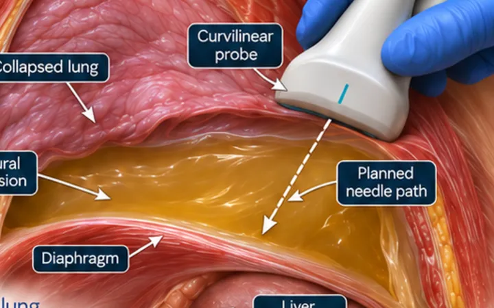
Thoracentesis: mark the pocket
A safe pleural tap needs three things seen at the bedside, not on the X-ray. Find the diaphragm, find the deepest pocket that stays clear of lung, liver and spleen through the whole breathing cycle, measure skin to fluid, and enter just above the rib below without moving the patient.
Airway
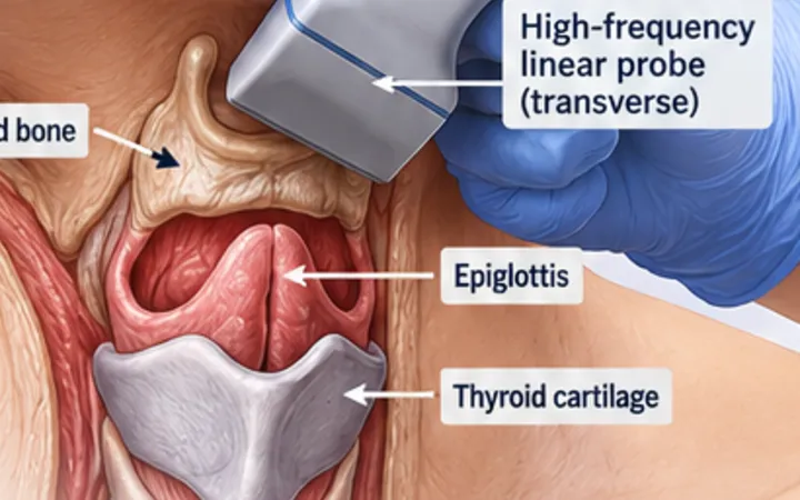
Epiglottis: find the swollen leaf
In a calm adult with a severe sore throat, a transverse scan above the thyroid cartilage can show a thickened, rounded epiglottis. It supports the diagnosis; it never excludes it. Airway assessment and ENT come first, and nobody with airway compromise should be scanned.
Airway
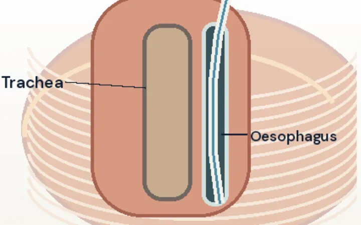
NG tube: ultrasound is a scout, not clearance
Ultrasound can show a nasogastric tube in the oesophagus on the left side of the neck, or in the stomach, which helps troubleshoot a difficult insertion. In UK practice it does not confirm position. Before anything goes down the tube, use aspirate pH 1 to 5.5 or a correctly interpreted X-ray.
Lung
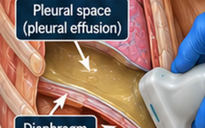
Pleural effusion: simple or septated?
Once you have found an effusion, look inside it. Black fluid can be a transudate or an exudate. Strands, septations, swirling echoes or loculated pockets mean an exudate, and in an unwell patient, possible pleural infection that may need a drain.
Lung
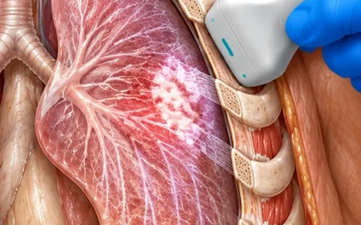
B-lines: map the pattern
B-lines are not a diagnosis. Map whether they are focal, patchy or diffuse, then look at the pleural line, spared areas and any small consolidation.
Airway
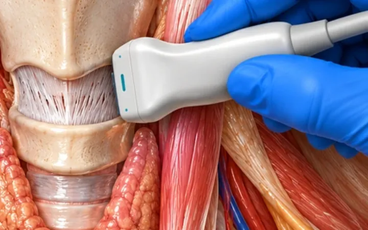
Cricothyroid membrane: find it before the emergency
When the neck landmarks are hard to feel, ultrasound can find the thyroid cartilage, the cricoid and the membrane between them. Find and mark it while the patient is oxygenating, never during a can't intubate, can't oxygenate emergency.
Lung
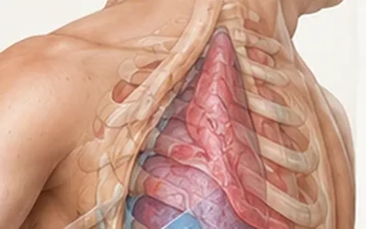
Consolidation: watch the air bronchograms move
Bright dots in consolidated lung are air in the airways. If they move outwards as the patient breathes in, the airway is open and pneumonia is likely. If they stay still, think about collapse.
Lung
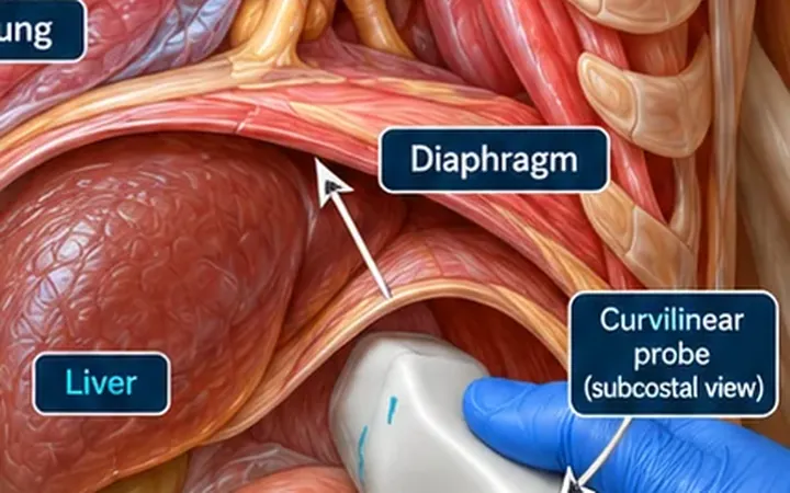
Diaphragm ultrasound: watch the sniff
A weak or paralysed diaphragm causes unexplained breathlessness and failure to wean. Put the probe below the ribs, ask for a sniff, and watch which way the diaphragm moves. Towards the probe is normal. Away from it is paradoxical.
Airway
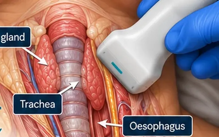
Intubation check: spot the double tract
Watch the neck as the tube goes in. One air shadow in the midline means the tube is in the trachea. A second air shadow beside it, where the oesophagus lies, means it is in the wrong place. Pair it with capnography, every time.
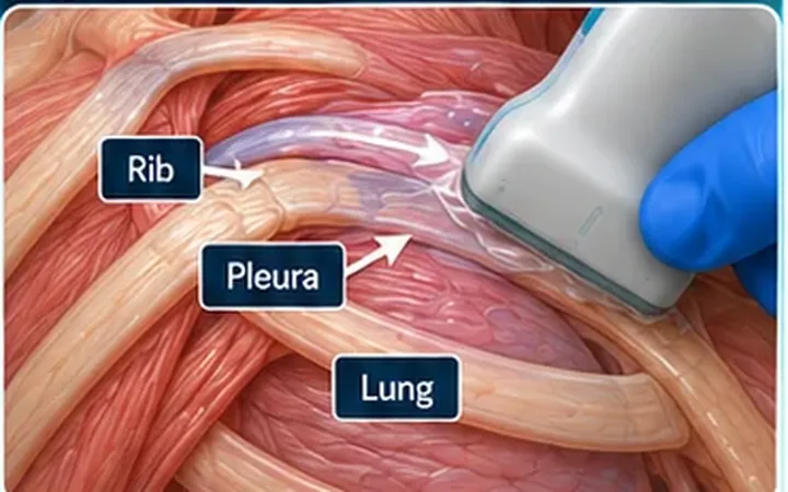
Lung
Lung: absent sliding is not enough
No sliding means look harder, not pneumothorax. B-lines or a lung pulse rule it out under the probe, and a lung point rules it in.
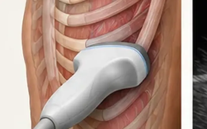
Lung
Pleural effusion: find the spine above the diaphragm
In a normal scan the spine stops at the diaphragm, because air-filled lung hides it. When fluid fills the base, the spine carries on above the diaphragm. Add black fluid and you have an effusion.
Abdomen and pelvis
Free fluid, the gallbladder and bile duct, kidneys, bladder, stomach, bowel and the pelvis, in adults and children.
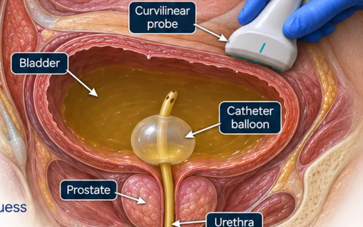
Renal
Blocked catheter: find the balloon
A catheter that stops draining is blocked, misplaced, or has no urine to drain. A scan above the pubic bone tells these apart in a minute. A full bladder around the balloon means blocked. An empty bladder means look upstream. No balloon in the bladder may mean it lies in the urethra.
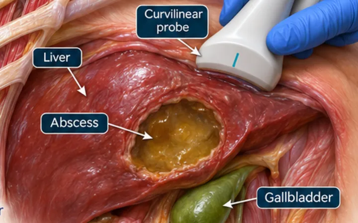
Abdomen
Liver abscess: fever and a dark round pocket
Fever and right upper quadrant pain with a gallbladder that does not explain them should make you sweep the whole liver. A round or irregular dark lesion with debris, no internal flow and bright tissue behind it suggests an abscess. An early abscess can look solid and bright, so a normal scan does not exclude it.
Renal
Stone at the bladder: look behind it
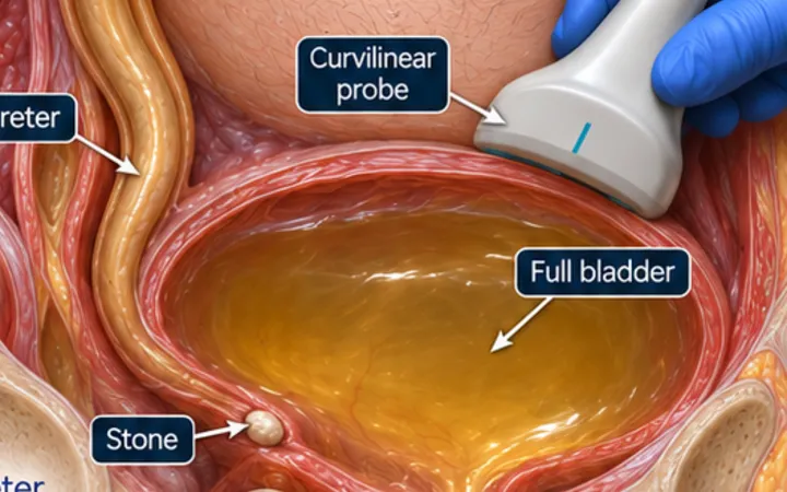
The junction of the ureter and the bladder is a common place for a stone to stick, and a full bladder gives a clear window onto it. A bright echo with a shadow or twinkle at the back of the bladder, with a dilated ureter or no jet on that side, suggests a stone. A normal scan does not exclude one.
Abdomen
Bowel ischaemia: read the wall
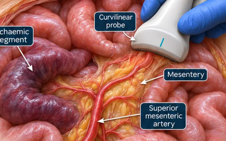
Severe abdominal pain out of proportion to the examination, especially in an older patient with atrial fibrillation or vascular disease, can be mesenteric ischaemia. Ultrasound can show a thick bowel wall with little flow, free fluid, or gas in the wall or portal veins. Early ischaemia can look normal, so a normal scan never excludes it, and CT angiography decides.
Pelvis
Adnexal ring: not always the ectopic
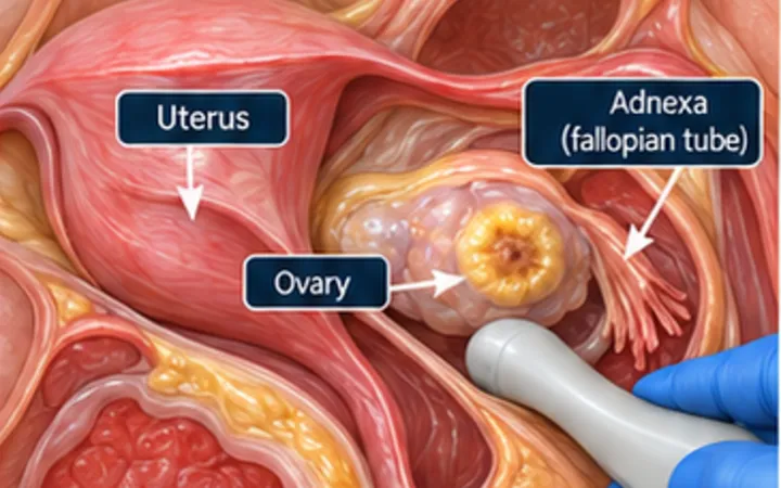
A ring in the adnexa with a positive pregnancy test is worrying, but the commonest ring is a normal corpus luteum. It sits inside the ovary; a tubal ectopic usually sits separate from it. Find the ovary first, then judge the ring, and never let the scan delay a referral.
Abdomen
Ascites: simple or complex?
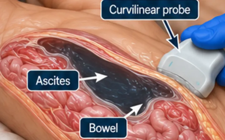
Simple ascites is black, free and moves with the patient. Debris, septations or loculated pockets point towards infection, blood or malignancy. But infected ascites often looks simple, so a black pocket never replaces a diagnostic tap.
Abdomen
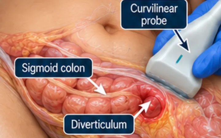
Diverticulitis: find the inflamed pouch
Scan exactly where it hurts. An inflamed diverticulum is a small dark pouch, often with a bright centre, arising from a thickened colon wall and surrounded by bright, inflamed fat. Look next for an abscess or free gas, which change management.
Abdomen
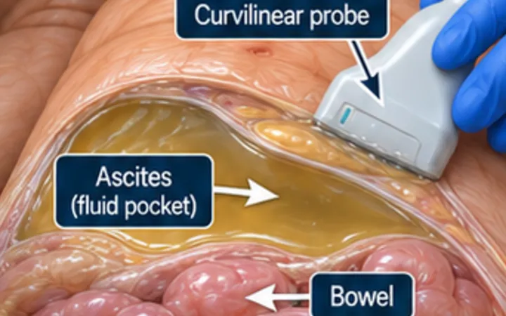
Paracentesis: pick the pocket
The safest place to drain ascites is the deepest pocket with no bowel and no vessel in the path, not the first black space you see. Sweep in two planes, check the wall with colour Doppler, measure the depth, and mark and drain without moving the patient.
Pelvis
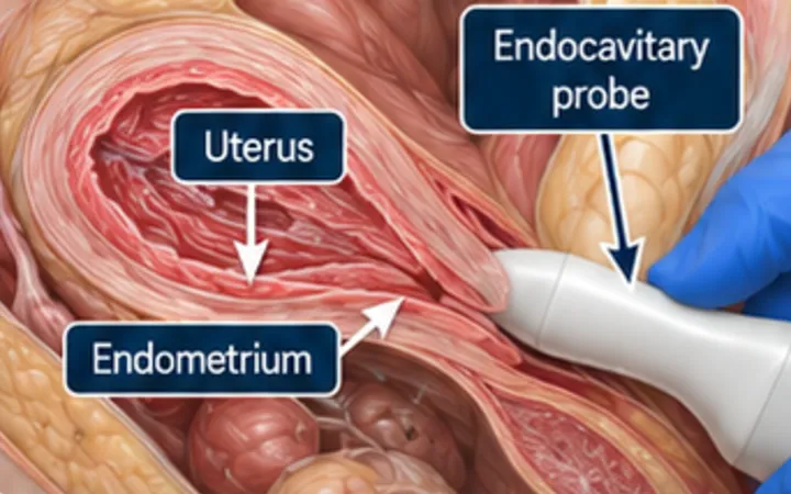
Retained products: thick and vascular
Bleeding after a miscarriage, termination or birth. A thick, uneven endometrium with blood flow inside it raises concern for retained products; clot is usually avascular. Thickness alone is not enough, and the scan never replaces the history, the hCG and gynaecology review.
Renal
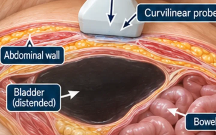
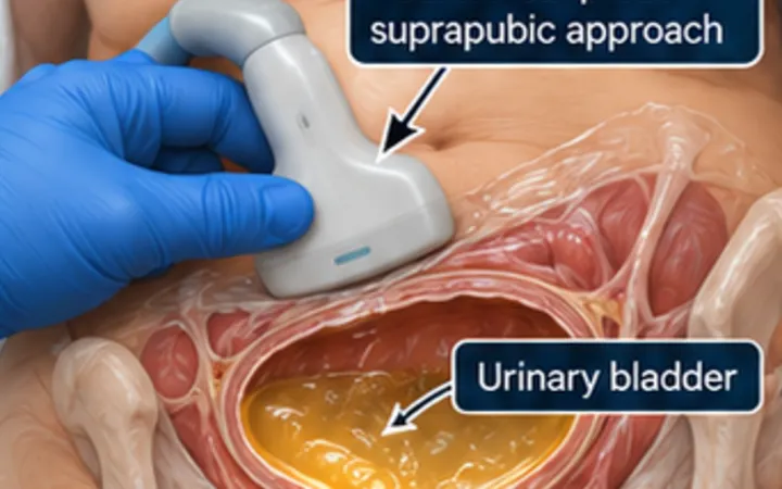
Suprapubic catheter: fill first, then find the window
A suprapubic catheter or bladder aspiration needs a full bladder and a clear path. A distended bladder pushes bowel away and gives a broad target. Confirm in two planes that no bowel lies between the skin and the bladder, and measure the depth.
Renal
Cauda equina: measure the residual
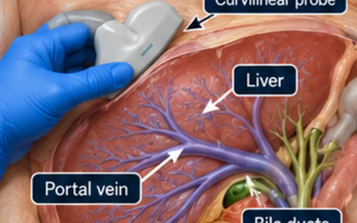
In suspected cauda equina syndrome, a bladder scan after the patient tries to void adds one objective sign. A large residual supports retention and raises concern. A normal residual does not exclude cauda equina syndrome, and the scan never replaces urgent imaging.
Abdomen
Liver air: portal venous gas or pneumobilia?
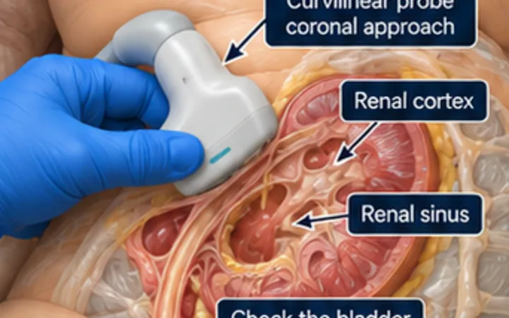
Bright gas echoes in the liver are either in the portal veins or in the bile ducts. Portal venous gas is carried out to the edge of the liver; pneumobilia stays central near the hilum. The difference matters, because portal venous gas can signal bowel ischaemia.
Renal
Pyelonephritis: a normal scan can be normal
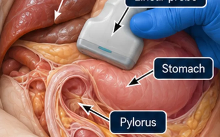
Most kidneys with pyelonephritis look normal on ultrasound, so the scan cannot rule the infection in or out. Its job is to find the complications which change management, above all an obstructed, infected kidney.
Abdomen
Pyloric stenosis: measure, then watch
A young baby with forceful, non-bilious vomiting. A thick, long pylorus that never opens supports pyloric stenosis. Measure the muscle and the channel, then watch for several minutes, because pylorospasm can look the same in a single frame.
eFAST
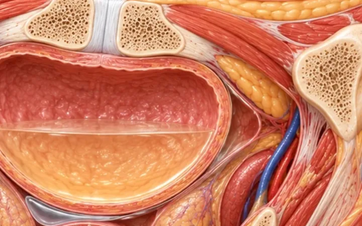
eFAST pelvis: sweep behind the bladder
The bladder is the window, not the target. Fan through the pelvis in sagittal and transverse so a small pocket of free fluid is not missed at the edge of one still image.
Abdomen
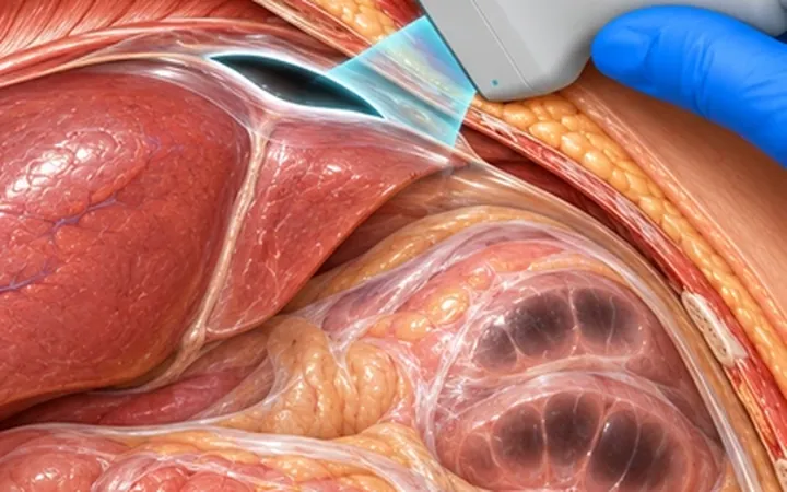
Pneumoperitoneum: inspect the stripe
Free air in the abdomen makes the peritoneal line under the abdominal wall unusually bright, with reverberation beneath it. Bowel gas sits deeper, under a normal thin stripe. A negative scan does not rule out perforation.
Renal
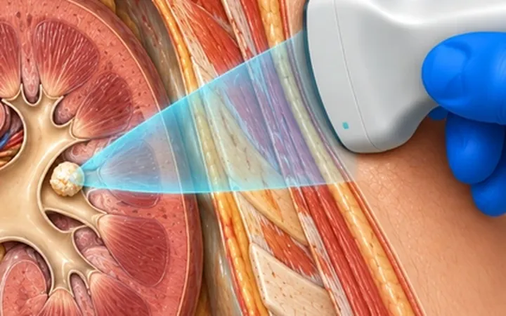
Renal stone twinkle: a clue, not a diagnosis
A flickering colour tail behind a bright focus can show a stone, but twinkle has false positives and depends on the settings. Confirm the bright focus, its shadow and the signs of obstruction before you call a stone.
Renal
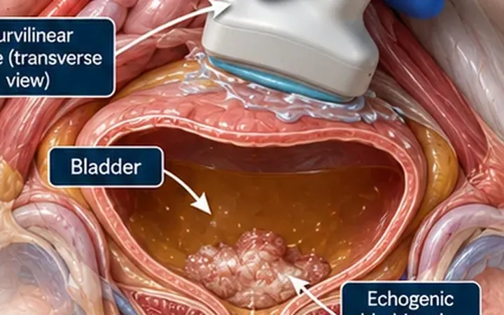
Bladder filling defect: move it, then add colour
Something bright sits in the bladder. Roll the patient and add colour Doppler. Clot moves and has no internal flow. A lesion stuck to the wall stays put and may show vessels.
Abdomen
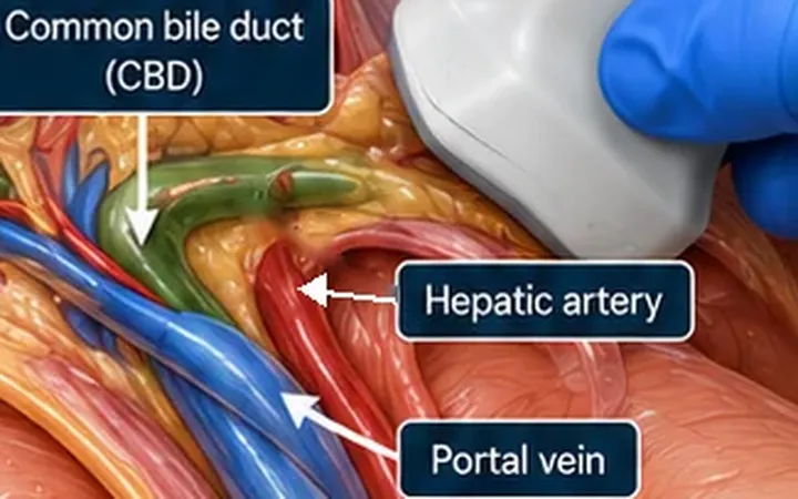
Common bile duct: find the parallel channel
Next to the portal vein lie two tubes, the hepatic artery and the bile duct. Add colour Doppler. The tube with no flow is the duct.
Pelvis
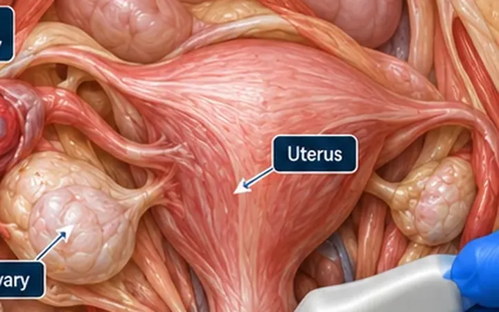
Ectopic pregnancy: empty uterus? Find the adnexa
A positive pregnancy test and pain. The bedside scan has one job first, to find a pregnancy inside the uterus. If you cannot, look beyond it, at the adnexa, the pouch of Douglas and the right upper quadrant.
Abdomen
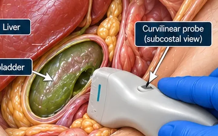
Gallbladder wall: thick does not equal cholecystitis
A gallbladder wall over 3 mm is a clue, not a diagnosis. Look for stones, focal tenderness, distension and pericholecystic fluid before you call cholecystitis.
Pelvis
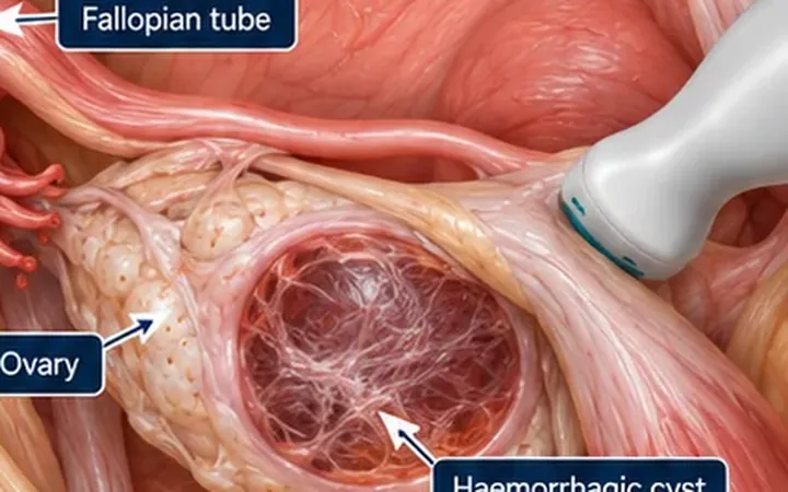
Haemorrhagic cyst: fishnet, not flesh
Lacy internal echoes and a clot with a concave edge and no flow inside it point to a haemorrhagic ovarian cyst. A solid part with blood flow does not, and needs formal imaging.
Pelvis
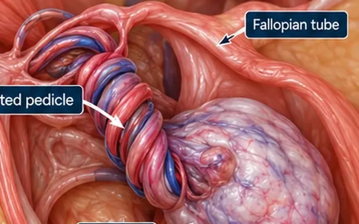
Ovarian torsion: follow the pedicle
Normal Doppler flow does not exclude torsion. Look for an enlarged ovary with peripheral follicles, and follow the vascular pedicle for a whirlpool. Clinical suspicion drives the referral.
Pelvis
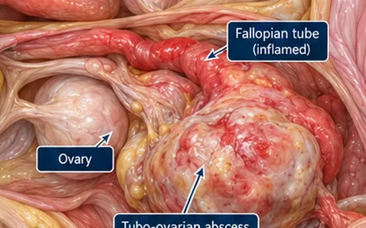
Tubo-ovarian abscess: think complex, thick and tender
Pelvic pain with fever and a complex, thick-walled, tender adnexal mass should raise concern for a tubo-ovarian abscess. POCUS raises the alarm. Formal imaging and gynaecology confirm it.
Renal
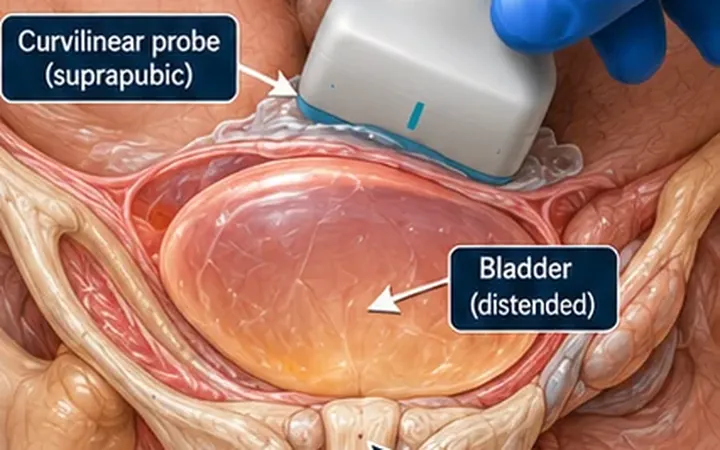
Urinary retention: measure the bladder, then look upstream
Measure the bladder in two planes to confirm it is full. Then scan both kidneys, because a large bladder with hydronephrosis means the obstruction has reached the upper tracts.
Abdomen
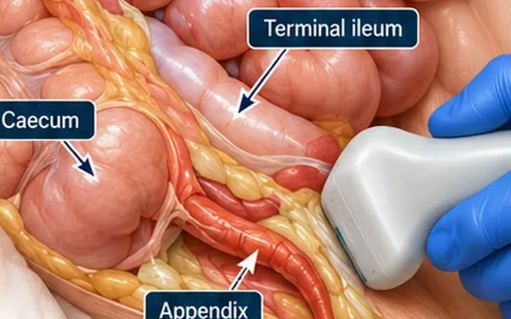
Appendix ultrasound: a positive scan helps
A blind-ending, non-compressible tube over 6 mm across in the right lower quadrant supports appendicitis. Not seeing the appendix does not exclude it. Learn graded compression, what to measure, and how to act on each result.
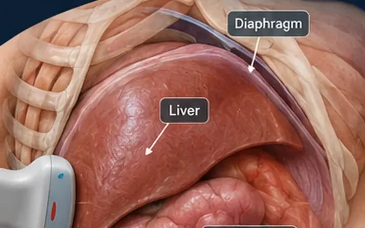
eFAST
eFAST: look at the liver tip first
In the supine patient, free fluid in the right upper quadrant often collects first at the inferior tip of the liver. Scan the whole view, not just Morison's pouch.
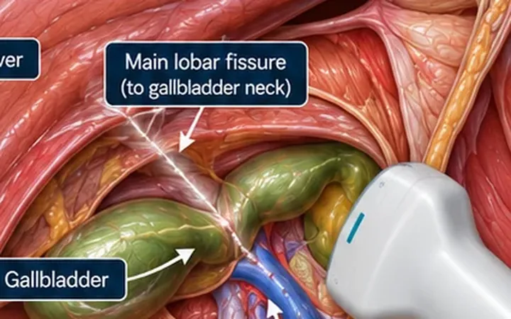
Abdomen
Gallbladder: follow the main lobar fissure
Can't find the gallbladder? Find the portal vein, then follow the bright line running from it. The main lobar fissure leads straight to the gallbladder neck.
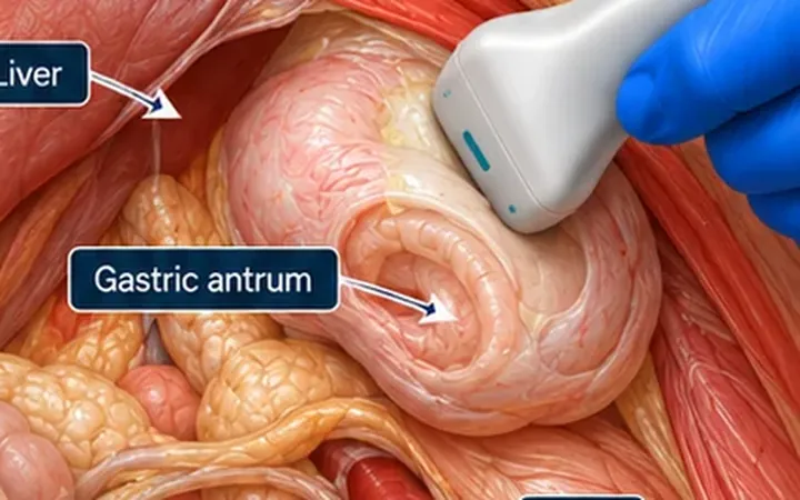
Abdomen
Gastric POCUS: empty, fluid or solid?
Before sedation, one view of the gastric antrum tells you whether the stomach is empty, holds clear fluid, or holds solid food. Answer that question first.
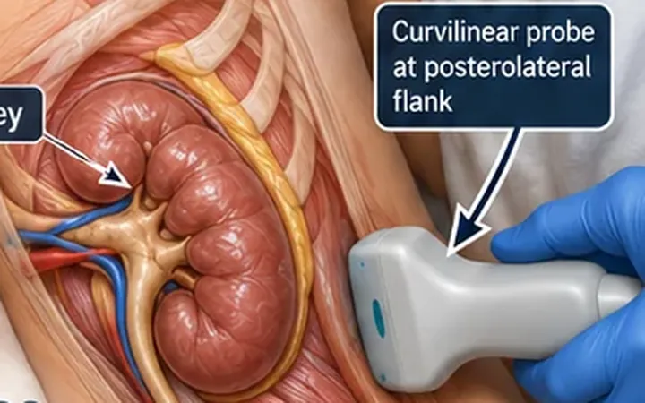
Renal
Hydronephrosis: black that branches
True hydronephrosis forms connected, branching black spaces running from the renal pelvis into the calyces. Cysts stay round and separate. Sweep the whole kidney before you decide.
Abdomen
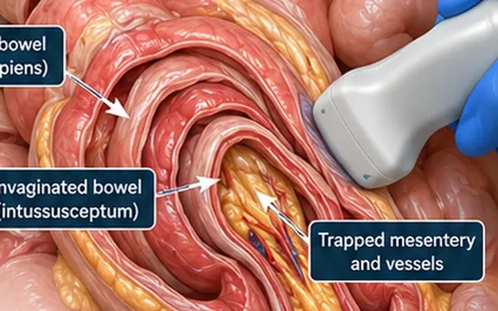
Intussusception: target, then sandwich
A child with colicky pain, drawing up the legs and periods of calm needs a look at the right side of the abdomen. A target in transverse and a sandwich in long axis make the diagnosis. Two planes, one answer.
Renal
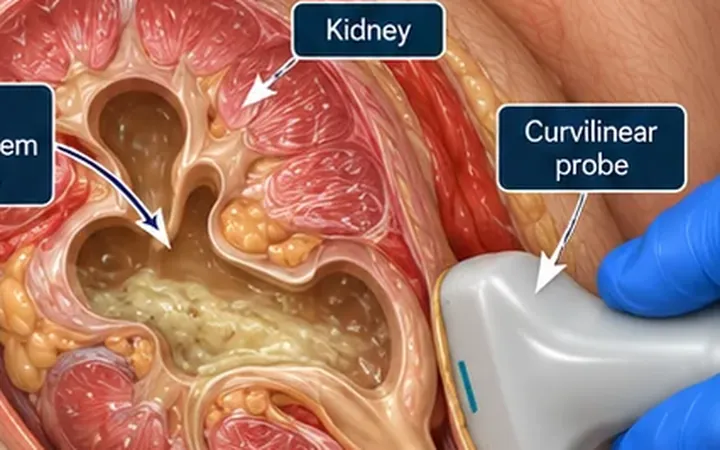
Pyonephrosis: hydronephrosis with dirty echoes
Hydronephrosis in a septic patient is an obstructed, infected kidney until proven otherwise. Debris, layering echoes or a fluid-debris level in the collecting system should make you think of pus and call urology now.
Renal
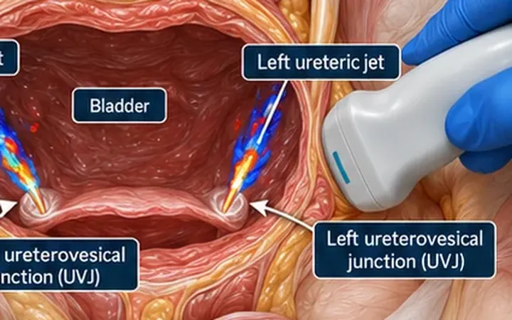
Renal colic: listen with colour
A ureteric jet is urine squirting into the bladder. Colour Doppler shows it. A jet missing on the painful side, with hydronephrosis on that side, supports an obstructing stone. Wait, watch and compare both sides.
Abdomen
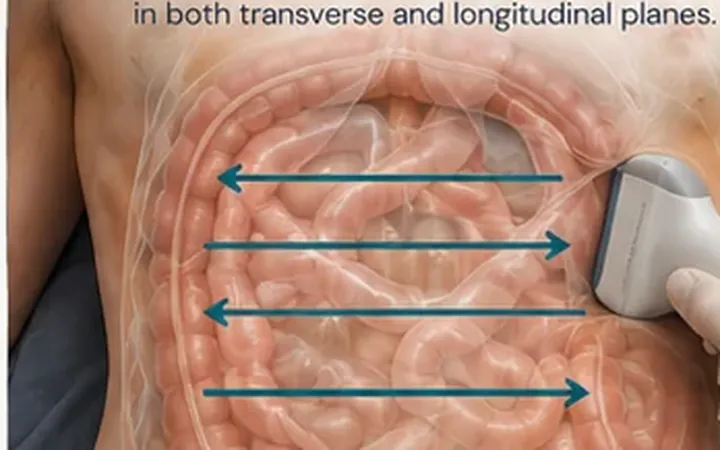
Small bowel obstruction: follow the to-and-fro
Dilated, fluid-filled small bowel with contents moving back and forth without getting anywhere suggests mechanical obstruction. Follow the dilated loops until they collapse to find the transition point.
Soft tissue and joints
Collections, foreign bodies, tendons and joint effusions.
Soft tissue
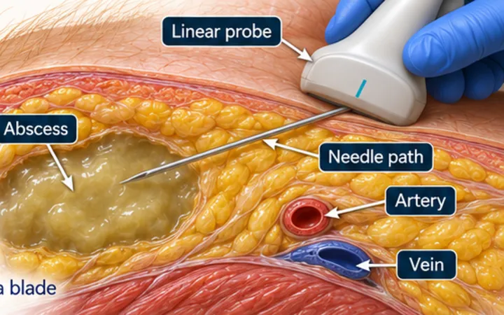
Abscess drainage: plan the needle path
Before any needle or blade, ultrasound measures how deep the pocket starts and how wide it is, shows the vessels nearby and finds the shortest safe route. Colour Doppler comes first, because a pseudoaneurysm can look like an abscess. Then mark the skin or watch the needle, and rescan for a remaining pocket.
Soft tissue
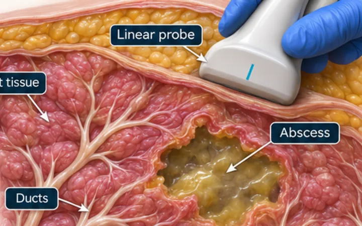
Breast abscess: aspirate before you cut
In a hot, tender breast, ultrasound separates mastitis from a collection. A dark or complex pocket with no internal flow suggests an abscess, and oedema without a pocket suggests mastitis. Most abscesses are treated by needle aspiration under ultrasound, repeated if needed, with antibiotics and a breast team. A red breast that does not settle needs the breast clinic.
Soft tissue
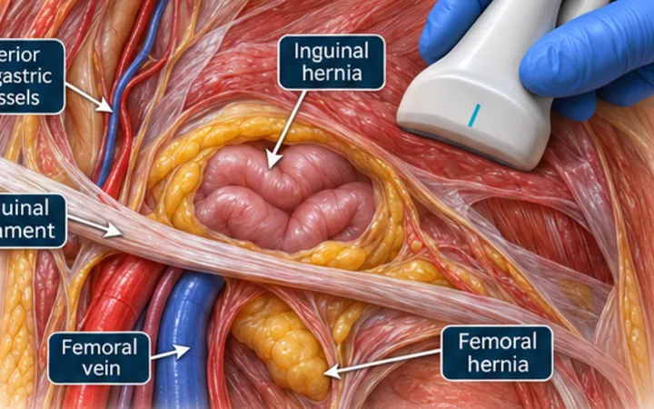
Groin lump: inguinal or femoral?
Inguinal hernias lie above the inguinal ligament. Femoral hernias lie below it, beside the femoral vein, and often strangulate. Scan along the ligament and over the femoral vessels, at rest and on straining. Colour Doppler every groin lump, because a saphena varix also grows with straining, and a pseudoaneurysm can look like an abscess.
Soft tissue
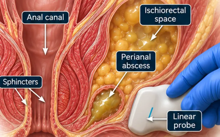
Perianal abscess: judge the depth
A small lump beside the anus can be the top of a much deeper collection. Ultrasound from the skin notes the depth of the pocket and how it relates to the sphincter. A collection tracking deep needs imaging and surgery. Ultrasound can miss deep and supralevator collections, and bright gas with dirty shadowing raises concern for Fournier gangrene.
Soft tissue
Strangulated hernia: read the sac
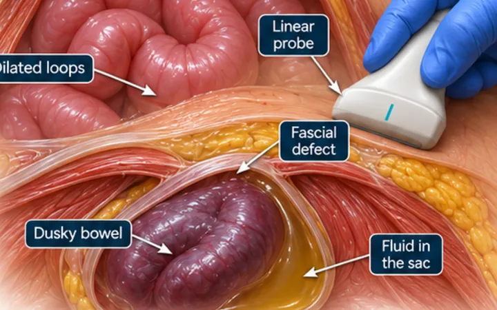
In a painful hernia that will not go back, ultrasound looks for bowel at risk. A thick wall, fluid in the sac, no peristalsis and little flow in the wall suggest strangulation, and dilated loops upstream suggest obstruction. Preserved flow does not exclude it, so a painful, irreducible hernia goes to the surgeons whatever the scan shows.
Soft tissue
Parotid swelling: find the collection
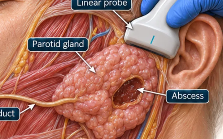
A hot, tender swelling in front of the ear is usually acute parotitis, which settles with fluids, antibiotics and sialogogues. Ultrasound finds the minority with a collection: a dark, complex pocket inside the gland with flow in its rim. The facial nerve runs through the gland, so drainage belongs to ENT, and the deep lobe hides behind the jaw.
MSK
Clavicle: follow the bright line
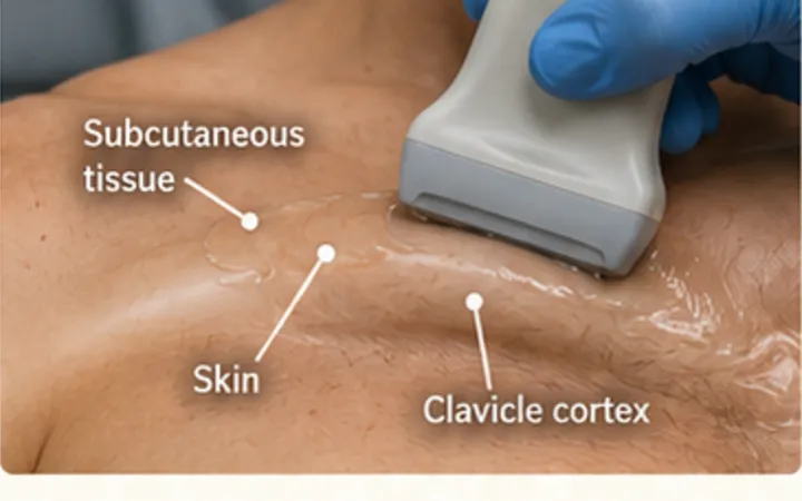
The clavicle lies just under the skin, so its cortex is easy to follow from the sternum to the shoulder. A step, a gap or a haematoma over the bone supports a fracture. It is especially useful in children. Check the skin, the nerves and the circulation, and remember the ends of the bone.
Soft tissue
Dental swelling: find the vestibular pocket
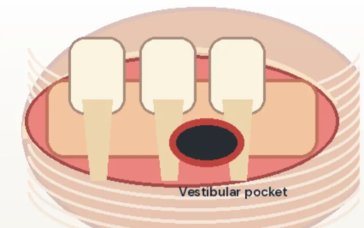
Facial swelling from a tooth can be cellulitis or a collection. Over the swelling, a hypoechoic pocket beside the bright jaw bone means pus that may drain. Ultrasound does not see the deep neck spaces, so trismus, drooling or floor-of-mouth swelling put the airway first.
MSK
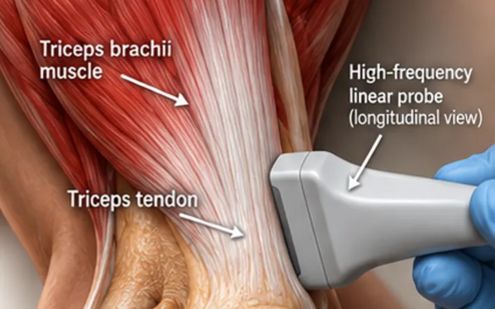
Distal triceps: find the olecranon attachment
Weak elbow extension after a fall onto an outstretched hand can be a triceps rupture. Follow the triceps tendon to the olecranon. A gap, a retracted stump or a small bony flake pulled off the olecranon suggests a tear. Swelling can hide it, so look in two planes and test extension.
Soft tissue
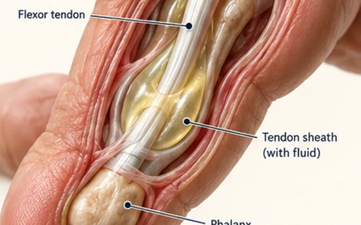
Finger infection: fluid in the sheath
A red, swollen finger may be cellulitis, a superficial abscess or infection inside the flexor tendon sheath. Follow the flexor tendon: fluid around it within a distended sheath supports flexor tenosynovitis, a hand surgery emergency. The scan supports the Kanavel signs; it does not replace them.
MSK
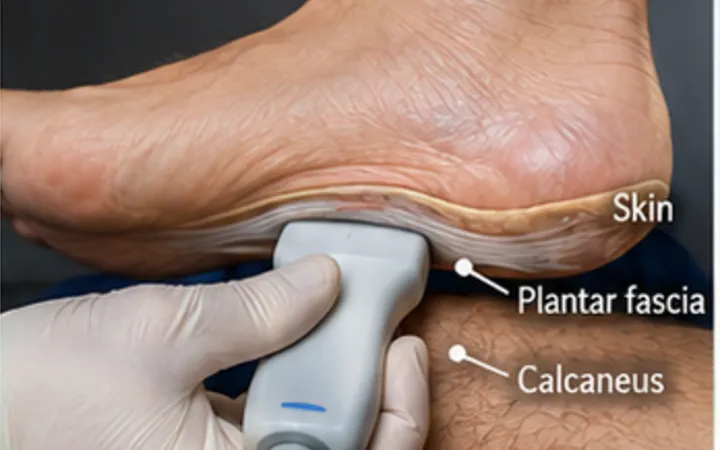
Heel pain: thick or torn?
Start at the calcaneus and follow the plantar fascia forwards. A fascia thicker than about 4 mm, dark and losing its fibres near the heel suggests plantar fasciitis. A gap with fluid suggests a rupture, often after a sudden pop or a steroid injection.
Soft tissue
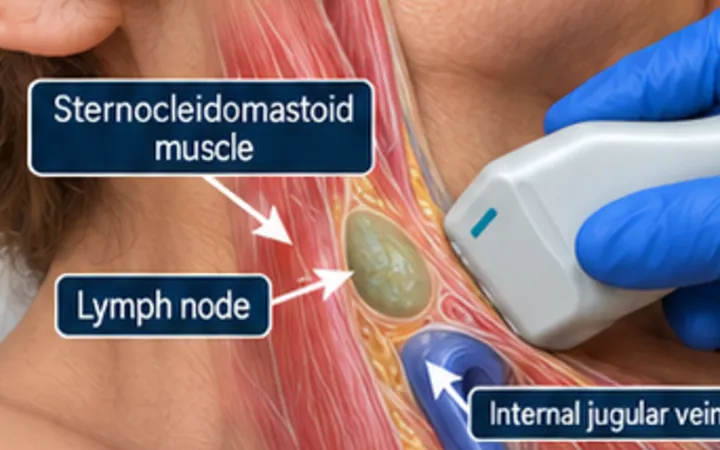
Neck lump: hilum or pus?
A tender neck lump is usually a node. An oval lump with a bright central hilum is a lymph node; a round collection with no hilum, debris and posterior enhancement suggests an abscess. A necrotic node can look just like an abscess, so use the whole picture.
MSK
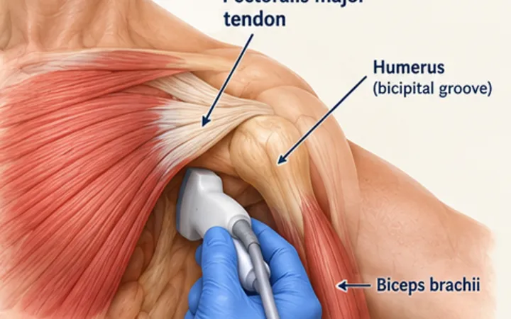
Pectoralis major tear: look beyond the shoulder
A pop at the front of the shoulder during a bench press, then bruising down the arm and a thin anterior axillary fold. Follow the pectoralis major tendon to its insertion on the humerus. A gap or a retracted stump suggests a tear, and complete tears do best with early repair.
Soft tissue
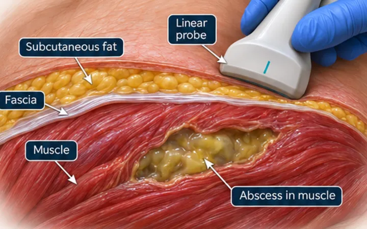
Pyomyositis: look below the fascia
Deep, constant muscle pain with fever, often in the thigh, buttock or calf, can be pyomyositis, an infection inside the muscle. Scan past the fat into the muscle. Swollen, disorganised muscle or a complex pocket within it is the clue. Early on it can look almost normal, so MRI and surgical review follow.
MSK
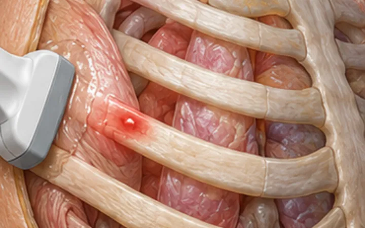
Rib fracture: trace the cortex
Put the probe on the most tender point and slide along the rib. A step, a gap or a haematoma on the bright cortex supports a fracture, often one an X-ray missed. Then look under the rib for the pleura, because the complications matter more than the crack.
MSK
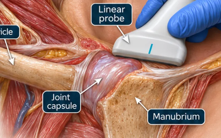
Sternoclavicular joint: look for capsular lift
A hot, swollen sternoclavicular joint can be septic arthritis, especially in people who inject drugs, have diabetes or a central line. Ultrasound across the joint shows fluid lifting the capsule. Compare both sides. Fluid does not prove infection, and spread behind the joint needs CT or MRI.
MSK
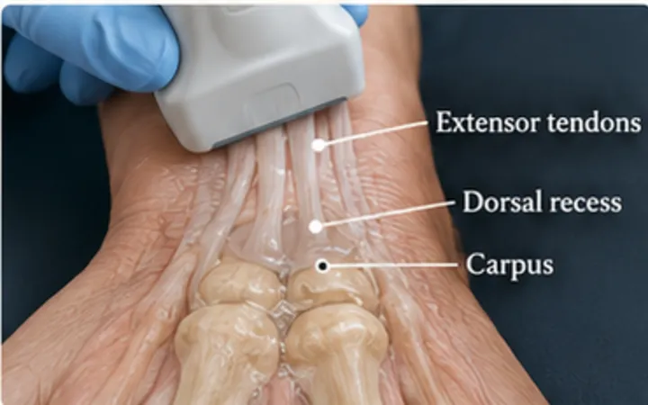
Wrist effusion: find the dorsal recess
A hot, swollen wrist could be a joint effusion, tenosynovitis, a ganglion or cellulitis. Joint fluid distends the dorsal recess over the carpal bones, deep to the extensor tendons. Find the layer, compare the other side, and aspirate a joint that might be infected.
MSK
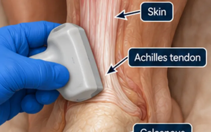
Achilles lump: xanthoma or tear?
Not every thick Achilles is torn. A xanthoma thickens the tendon but keeps its fibres continuous, often on both sides, and points to inherited high cholesterol. A tear breaks the fibres and leaves a gap which opens with gentle dorsiflexion.
MSK
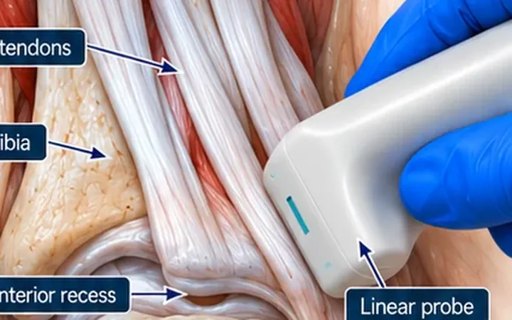
Ankle effusion: open the anterior recess
Ankle joint fluid collects in the anterior recess, deep to the extensor tendons, between the tibia and the talus. Plantarflexion opens the recess and makes a small effusion easier to see and to aspirate.
MSK
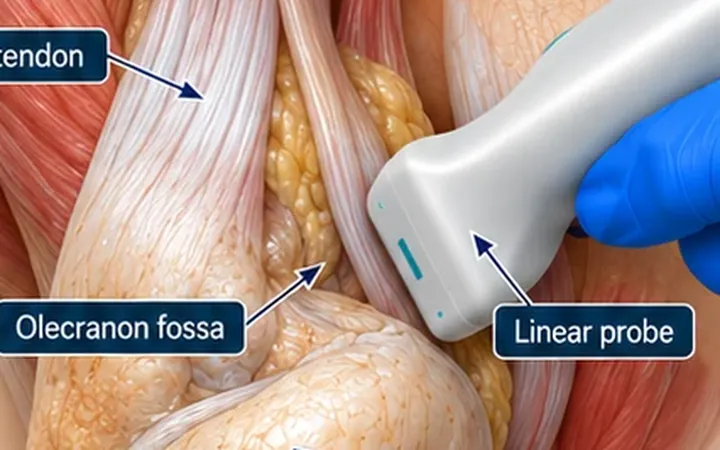
Elbow effusion: lift the fat pad
Fluid in the elbow joint collects in the posterior recess and lifts the posterior fat pad out of the olecranon fossa. Ultrasound shows the effusion well, but it may not show the fracture which caused it.
MSK
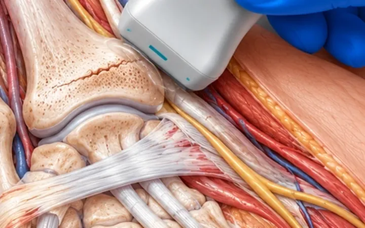
Fracture ultrasound: follow the cortex
Bone cortex is a bright, continuous line with a shadow beneath it. A step, gap or buckle at the point of tenderness becomes convincing when you see it again in a second plane.
Soft tissue
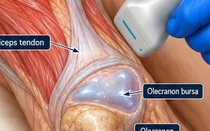
Olecranon bursa: keep the fluid in the right layer
Olecranon bursitis is fluid on top of the olecranon and the triceps insertion, not in the elbow joint. Find the layer first, then decide whether it is simple or complex. Ultrasound cannot tell you whether the bursa is infected.
MSK
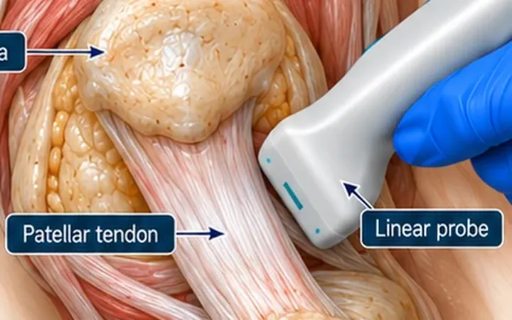
Patellar tendon rupture: follow it to the tibial tubercle
A complete patellar tendon rupture leaves a gap filled with fluid or haematoma between the patella and the tibial tubercle. Follow the tendon all the way to bone, and match the scan to the patient's ability to straighten the knee.
MSK
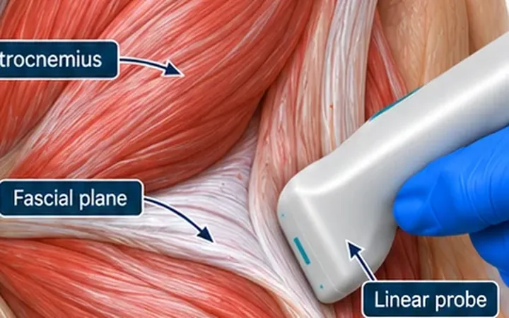
Tennis leg: look between gastrocnemius and soleus
A sudden pain in the medial calf during sport is often a tear of the medial gastrocnemius, with fluid tracking in the plane between it and soleus. It mimics a DVT, and a DVT can coexist, so the veins still need checking when the story fits.
Soft tissue
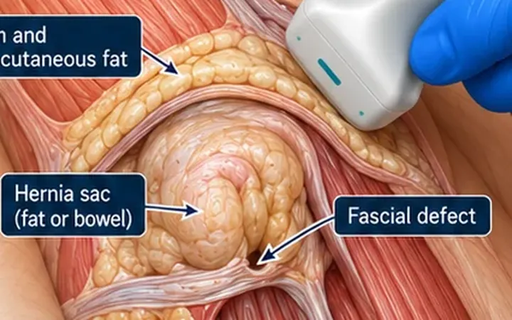
Abdominal wall hernia: stand them up and make it cough
A small hernia hides when the patient lies still. Scan the lump at rest, then during a cough, a strain or standing. Show the defect, what passes through it, and whether it goes back.
MSK
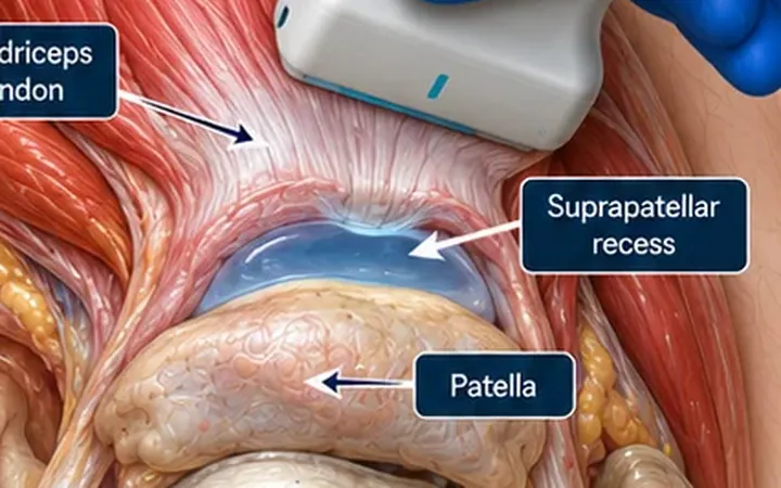
Hot knee: find the suprapatellar recess
Knee fluid collects in the suprapatellar recess, deep to the quadriceps tendon. Find it, squeeze fluid into it, and use it to guide aspiration. Ultrasound finds fluid. It does not tell you if it is infected.
MSK
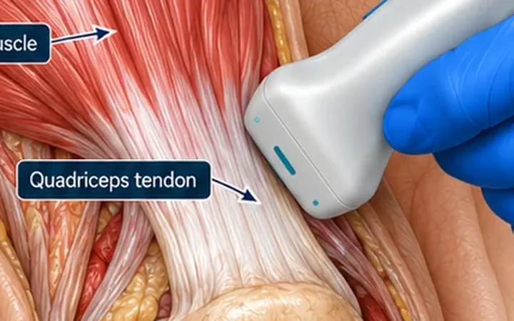
Quadriceps tendon: follow it to the patella
A fall, a giving-way knee and a patient who cannot straight-leg raise. Follow the quadriceps tendon down to the top of the patella and look for a gap. Bend the knee gently, and a true tear opens.
MSK
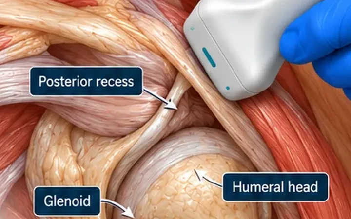
Shoulder effusion: start in the posterior recess
Glenohumeral fluid shows best behind the joint. From the posterior view, look for a dark space lifting the capsule off the humeral head, and compare with the other shoulder.
Soft tissue
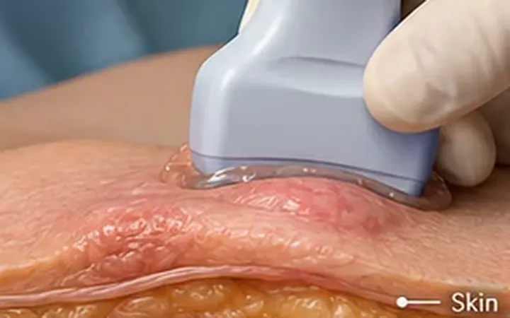
Abscess: make it swirl
An abscess can look grey and solid on a still image. Press gently with the probe. If the contents swirl, it is fluid. Then check colour Doppler before anyone reaches for a needle.
MSK
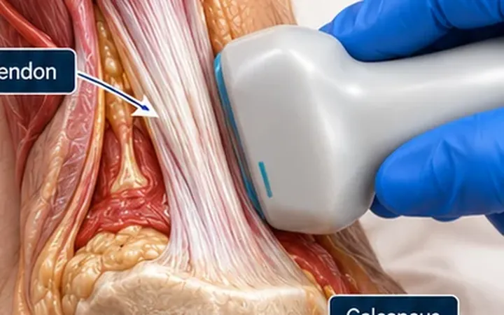
Achilles rupture: scan long, squeeze the calf
A still image of a torn Achilles tendon can look confusing. Scan in long axis, then squeeze the calf or move the ankle while you watch. If the movement stops at a gap, the tendon has ruptured.
MSK
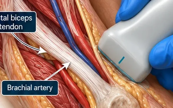
Distal biceps rupture: follow the tendon to the tuberosity
A pop at the elbow while lifting, then weakness and bruising. The tendon can look intact in the elbow crease when it has torn from the bone. Follow it all the way to the radial tuberosity. An empty insertion is the clue.
Soft tissue
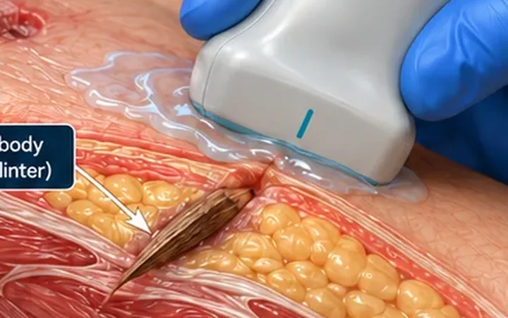
Foreign body: bright, then read the shadow
X-ray misses wood, plastic and thorns. Ultrasound sees every foreign body as a bright structure, and what lies behind it hints at the material. Find it, map it, then cut.
MSK
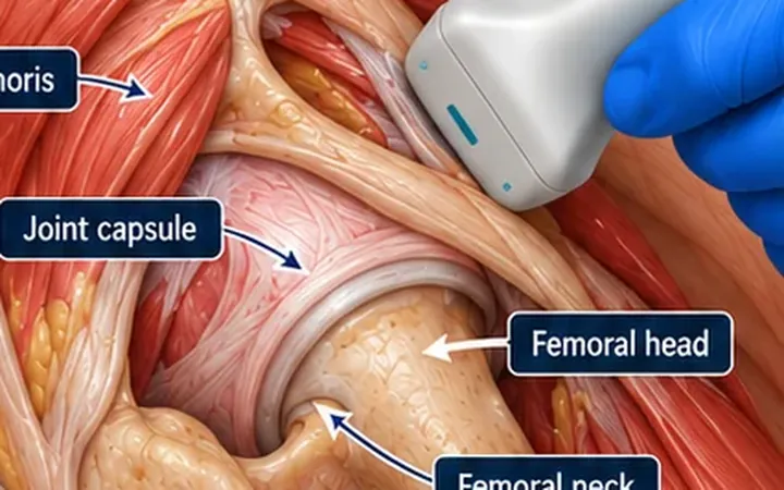
Hip effusion: compare the capsule distance
A limping child or a hot, painful hip raises the question of fluid in the joint. Align the probe with the femoral neck, measure the capsule-to-neck distance, then scan the other hip. Asymmetry is the strongest sign.
Soft tissue
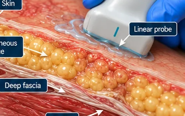
Necrotising fasciitis: look below the cobblestones
Cobblestoning fits cellulitis. Fluid along the deep fascia and gas in the soft tissue raise the alarm for necrotising fasciitis. Use the scan to escalate faster, never to reassure.
Soft tissue
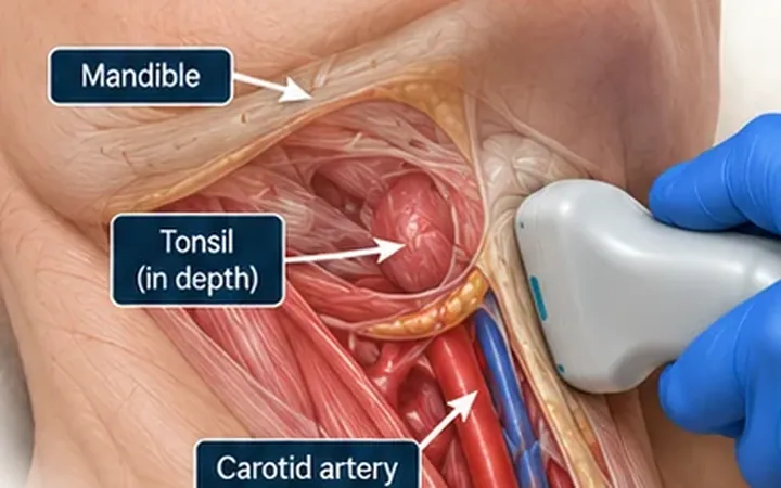
Peritonsillar abscess: scan from the neck
Trismus and gagging make an intraoral look difficult. A linear probe under the angle of the jaw shows whether there is a collection to drain, and where the carotid artery is before anyone reaches for a needle.
MSK
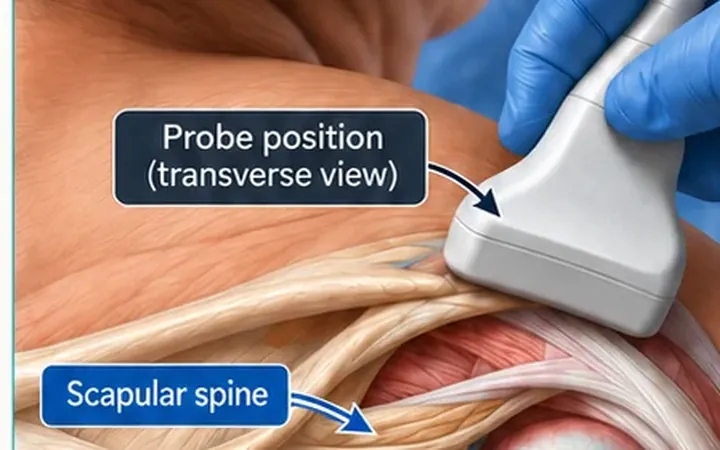
Shoulder: one posterior view, before and after reduction
Put the probe below the scapular spine and the glenoid and humeral head sit side by side. Where the head lies in relation to the glenoid tells you if the shoulder is out, which way, and if it has gone back in.
MSK
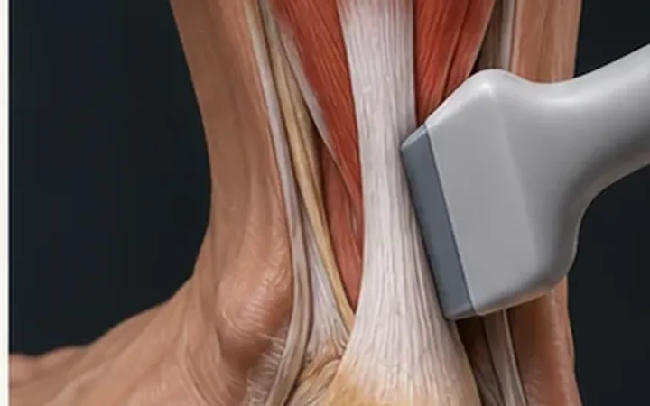
Tendon tear or anisotropy? Heel-toe before you call it
A normal tendon turns dark when the beam hits it at an angle. Rock the probe until the fibres brighten. If the dark area stays, think tear.
Eye and scrotum
Time-critical small parts, scanned safely.
Small parts
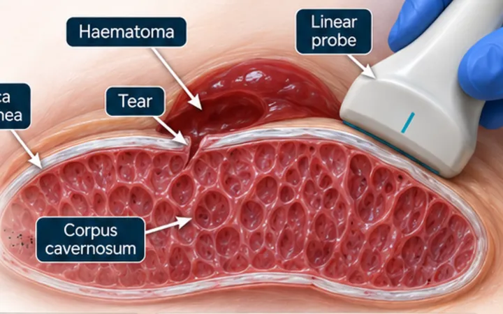
Penile fracture: find the tunica break
A snap, immediate loss of erection and a swollen, bruised penis are a penile fracture, a tear of the tunica albuginea. Ultrasound can show a break in the bright tunica with haematoma at the defect and locate it for the surgeon. A haematoma over an intact tunica is not proof, and a normal scan does not exclude a fracture: urgent urology either way.
Small parts
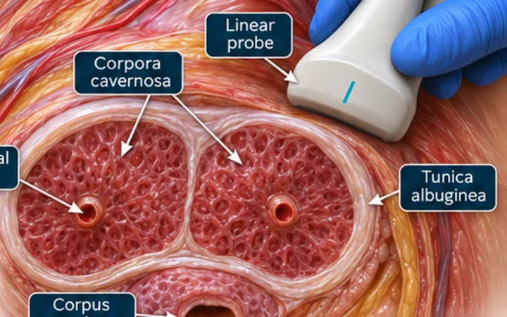
Priapism: read the cavernosal flow
A painful, rigid erection lasting over four hours is ischaemic priapism until proven otherwise, a urological emergency. Colour Doppler of the cavernosal arteries shows little or no flow in ischaemic priapism and preserved or high flow, sometimes with a turbulent blush, in the non-ischaemic type. The corporal blood gas decides, and neither test may delay treatment.
Eye
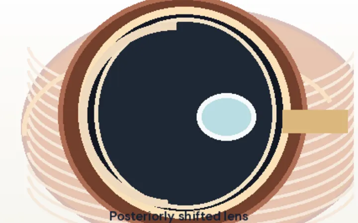
Eye trauma: has the lens moved?
After blunt eye trauma, find the lens first. Through the closed lid it sits just behind the iris as an oval with bright edges. A lens lying deep in the vitreous, or missing from its place, suggests dislocation. If the globe might be ruptured, do not scan: shield the eye and call ophthalmology.
Eye
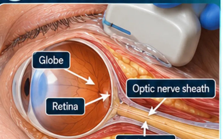
Optic nerve sheath: measure 3 mm behind the globe
Raised intracranial pressure distends the sheath around the optic nerve. Measure it 3 mm behind the globe, outer edge to outer edge, in two planes in each eye. A wide sheath is a clue that should speed up CT and senior review. It is not a diagnosis, and a normal value does not exclude raised pressure.
Small parts
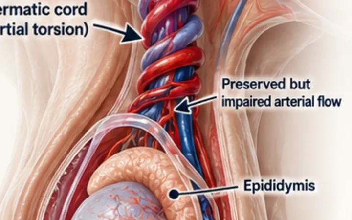
Partial testicular torsion: flow does not clear it
A partly twisted cord blocks the veins before the arteries, so colour flow can persist. Compare both testes, and where you can, look at the arterial waveform: lost or reversed flow in diastole suggests high resistance. Follow the cord for a twist. None of this replaces urgent urology review.
Small parts
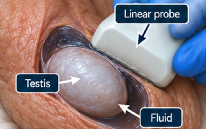
Scrotal fluid: clear or complex?
Fluid around the testis is common. Clear, black fluid is a hydrocele; debris, strands or septations suggest pus or blood. Either way, the fluid is not the diagnosis: look at the testis inside it and follow the cord.
Small parts
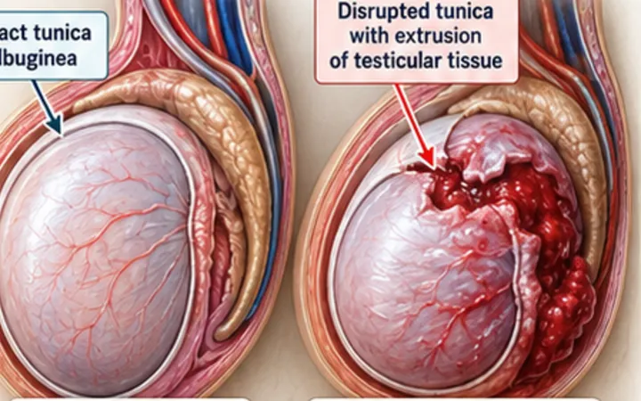
Scrotal trauma: follow the tunica
After a blow to the scrotum, the question is whether the testis has ruptured. Follow the bright tunica albuginea all the way round. A break in that line, with an irregular outline or tissue pushing through, suggests rupture, which needs surgery within hours. A bruise inside an intact tunica usually does not.
Small parts
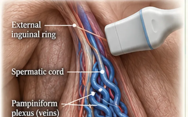
Varicocele: veins that wake up with Valsalva
A "bag of worms" above the testis is usually a varicocele: dilated veins of the pampiniform plexus that grow and fill with colour when the patient strains or stands. Most are on the left. A new right-sided varicocele, or one that does not empty lying down, needs a look further up.
Small parts
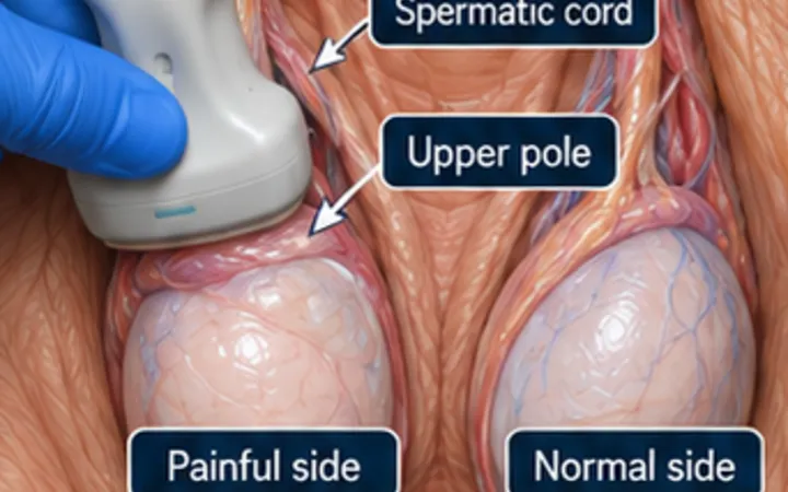
Appendix testis torsion: find the tender nub
A twisted appendix testis is a common cause of a painful scrotum in boys. It shows as a small, avascular nodule at the upper pole beside a testis with normal flow. It is a mimic, so the cord must still be followed, and urology must never wait for the scan.
Eye
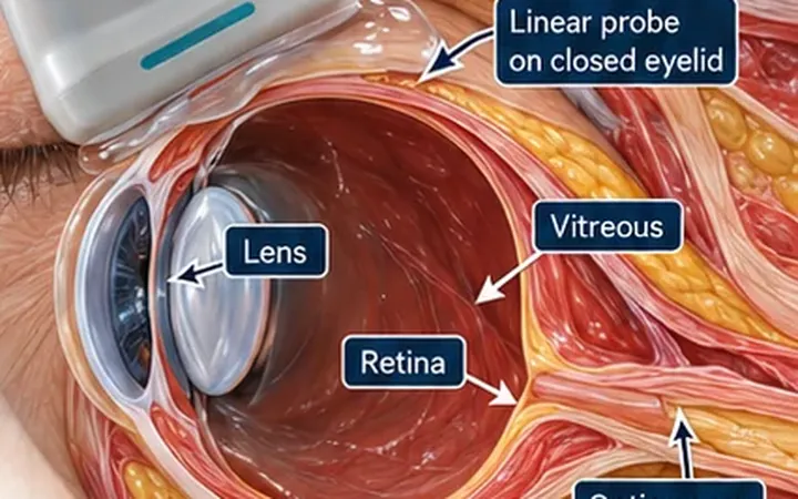
Ocular ultrasound: make the eye move
Sudden floaters, flashes or loss of vision raise three questions: retinal detachment, vitreous detachment or vitreous haemorrhage? A still image misleads. Ask the patient to move the eye and watch how the membrane behaves.
Small parts
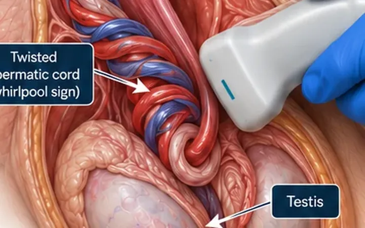
Testicular torsion: follow the cord
Torsion is a clinical diagnosis and urology comes first. When you do scan, do not stop at the colour box over the testis. Follow the spermatic cord and look for the twist, the whirlpool sign.
Nerve blocks
Ultrasound-guided regional anaesthesia for pain in the emergency department.
Nerve blocks
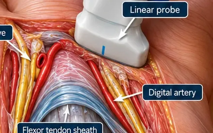
Digital nerve block: block the base, one deposit on each side
A digital nerve block numbs a finger for a laceration, a nail bed repair or a reduction. The palmar digital nerves run on each side of the flexor tendon sheath. Place a small deposit beside each one, and use ultrasound to see them.
Nerve blocks
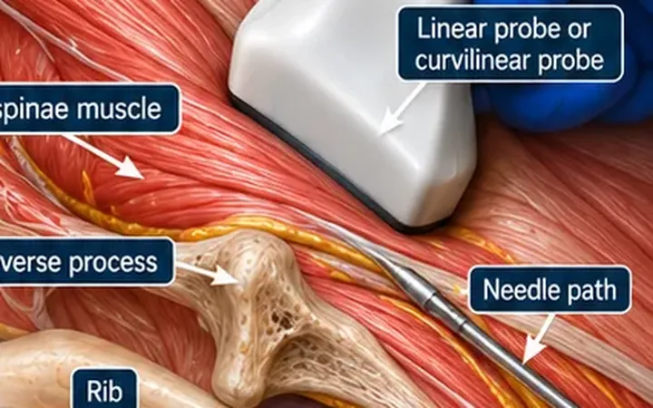
Erector spinae plane block: bone, muscle, then flood the plane
The erector spinae plane block places local anaesthetic on the transverse process, under erector spinae, far from the pleura. Find the flat-topped transverse process, lift the muscle off it and watch the fluid spread up and down.
Nerve blocks
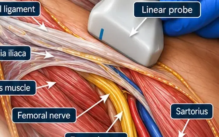
Fascia iliaca block, infra-inguinal: find the fascia, then flood the plane
The classic emergency department block for a fractured hip. Find fascia iliaca lying on iliacus, lateral to the femoral nerve, and fill the plane with a larger volume so it spreads across to the nerves of the thigh.
Nerve blocks
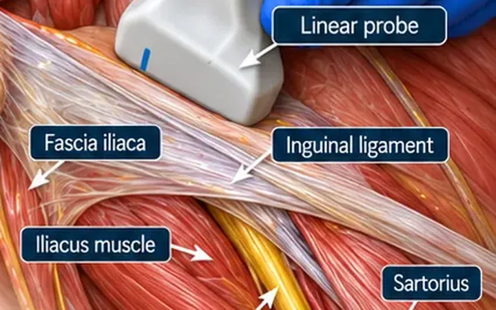
Fascia iliaca block, supra-inguinal: go above the ligament for better spread
The supra-inguinal approach places the local anaesthetic under fascia iliaca above the inguinal ligament, where it spreads up towards the lumbar plexus and reaches the lateral femoral cutaneous nerve more reliably. Find iliacus in a sagittal view near the anterior superior iliac spine and watch the fluid travel towards the head.
Nerve blocks
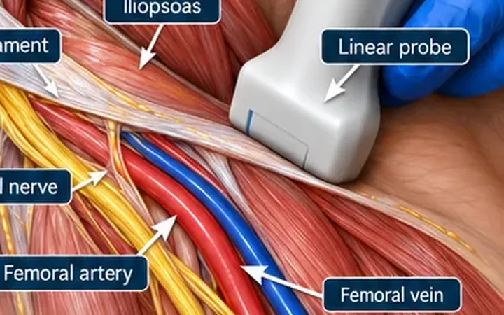
Femoral nerve block: artery first, nerve next, then wrap it
A femoral nerve block numbs the front of the thigh, most of the femur and the front of the knee. Find the femoral artery, slide lateral to the nerve under fascia iliaca, and watch the local anaesthetic surround it.
Nerve blocks
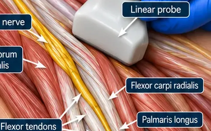
Median nerve block: find the fascicles, then build a soft halo
A forearm median nerve block numbs the palm on the thumb side and the index and middle fingers. Find the honeycomb nerve between the finger flexors in the mid to distal forearm, and surround it with a small volume.
Nerve blocks
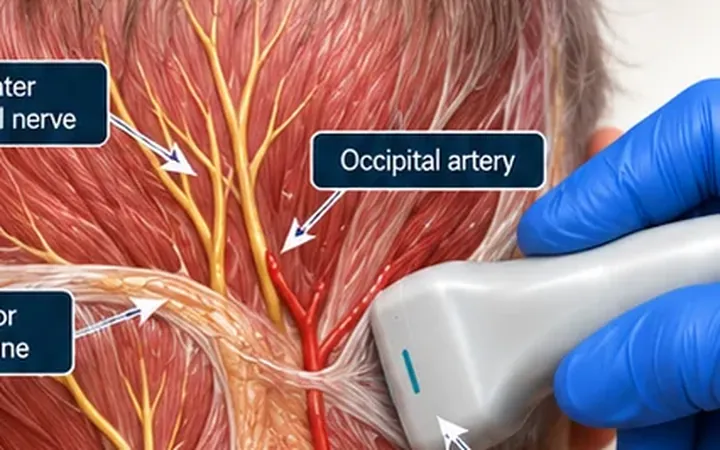
Occipital nerve block: follow the artery, then find the nerve
The greater occipital nerve runs beside the occipital artery at the back of the head. Colour Doppler finds the artery in seconds, which turns a blind injection into a targeted one. Rule out a dangerous headache first.
Nerve blocks
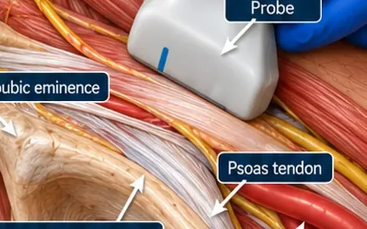
PENG block: find the ramus, then lift the tendon
The pericapsular nerve group block targets the small nerve branches to the front of the hip joint, where the psoas tendon crosses the pubic ramus. It relieves hip fracture pain on movement and tends to spare the quadriceps.
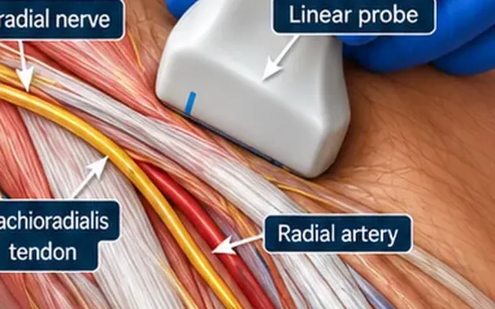
Nerve blocks
Radial nerve block: stay lateral, follow the nerve beside the artery
The superficial branch of the radial nerve supplies the back of the hand on the thumb side. In the forearm it runs beside the radial artery under brachioradialis. Use light pressure, find the artery, and look lateral for a small nerve.
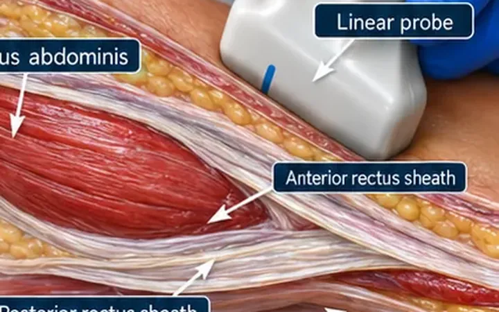
Nerve blocks
Rectus sheath block: behind the muscle, in front of the sheath
A rectus sheath block numbs the skin and muscle of the central abdominal wall. Place the local anaesthetic behind rectus abdominis, in front of the posterior sheath, on both sides for a midline wound.
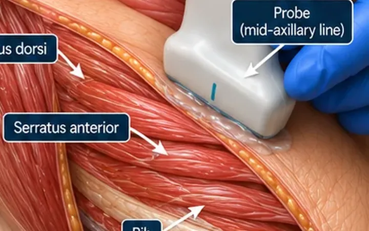
Nerve blocks
Serratus plane block: see the rib, see the pleura, then open the plane
Rib fractures hurt every time the patient breathes or coughs. A serratus anterior plane block numbs the side of the chest wall. Find the rib and the pleura first, and use the rib as your backstop.
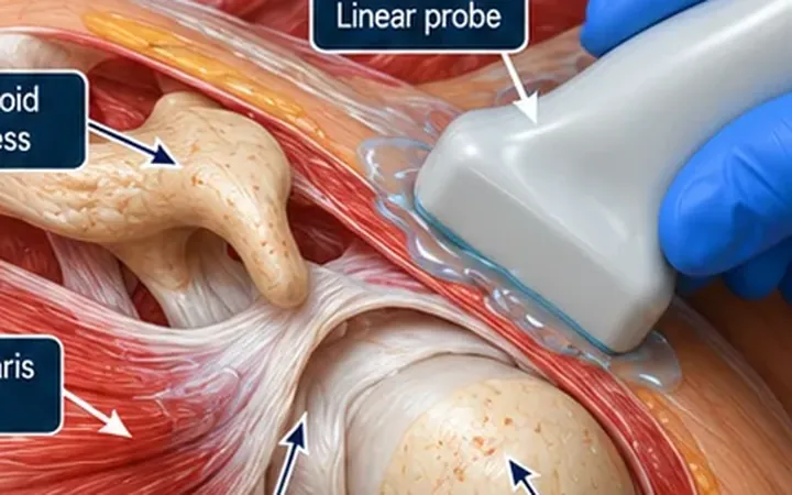
Nerve blocks
SHAC block: block the capsule, not the neck
The shoulder anterior capsular block places local anaesthetic on the front of the shoulder joint, away from the brachial plexus and the phrenic nerve. Stay lateral to the coracoid, find the subscapularis, and spread along the capsule.
Nerve blocks
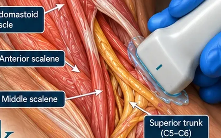
Shoulder reduction: low-volume superior trunk block
Trace the C5 and C6 roots down the neck until they join as the superior trunk, then surround it with a few millilitres of local anaesthetic. It numbs the shoulder for reduction with less risk to the diaphragm than an interscalene block.
Nerve blocks
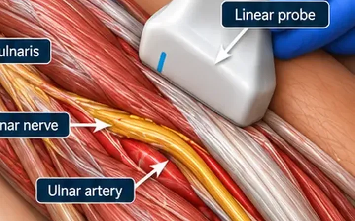
Ulnar nerve block: find the artery first, then block its neighbour
A forearm ulnar nerve block numbs the little finger side of the hand. Find the ulnar artery, the nerve lies on its ulnar side under flexor carpi ulnaris. Go a little higher in the forearm and the two separate.
Vascular access
Peripheral cannulation, central line safety and arterial lines with ultrasound.
Vascular access
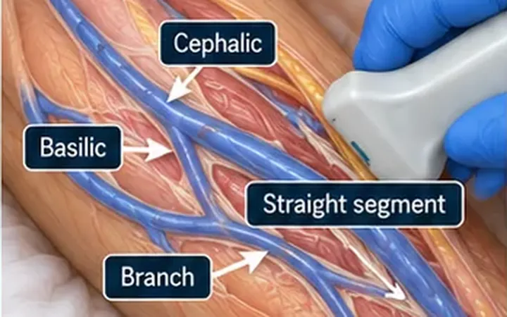
Choose the vein: pick the straight run
The best vein is not always the biggest. Survey the arm first, then choose a straight, compressible, superficial segment with enough length to hold the catheter, away from branches and valves.
Vascular access
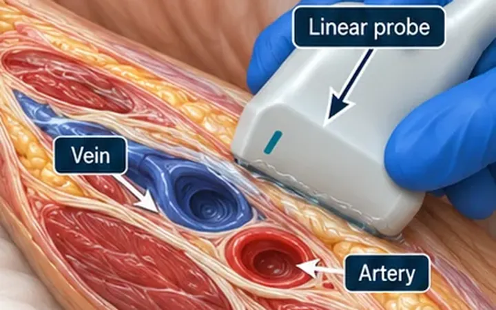
Vein or artery: compress first
Before you puncture, press gently on the target. A vein collapses and an artery stays open. Colour Doppler supports the answer, but colour depends on angle and settings, so do not trust it alone.
Vascular access
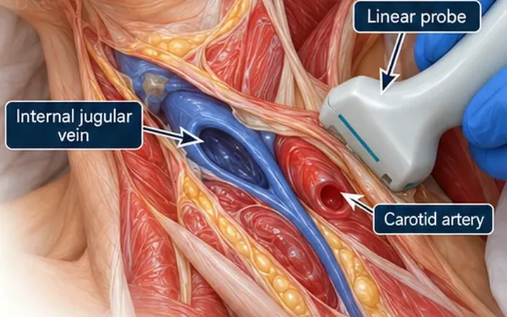
Central line: see the wire before you dilate
Flashback and easy wire passage do not prove the wire is in the vein. See the wire inside the internal jugular vein in two views before you dilate.
Vascular access
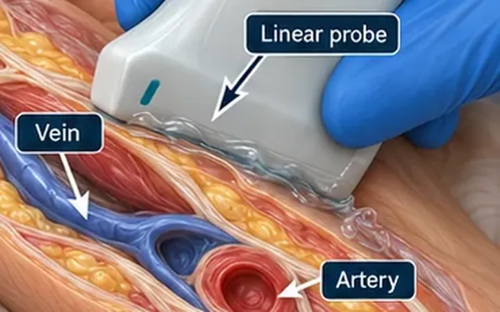
Peripheral access: centre the dot
Before you puncture, bring the vein to the middle of the screen, use the lightest pressure that keeps it round, and prove you can keep it there. A centred vein gives a straighter, safer needle path.
Vascular access
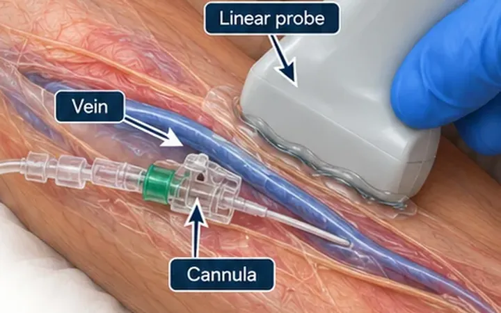
Flush test: saline swirl confirms the line
A small saline flush through the cannula, watched with a linear probe over the vein, tells you whether the fluid fills the lumen or the soft tissue. If the tissue swells, the line fails.
Vascular access
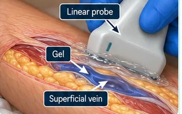
Light hand: do not collapse the vein
Veins hold low pressure, so a heavy probe flattens them and the target seems to vanish. Use plenty of gel and the lightest pressure that still gives a stable image.
Vascular access
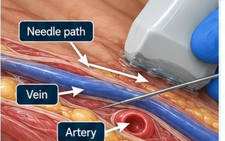
Posterior wall puncture: stop at the near wall
A flash of blood does not mean the needle is in the lumen. Slow down as the tip reaches the near wall, watch it enter the lumen, then stop before the back wall.
Vascular access
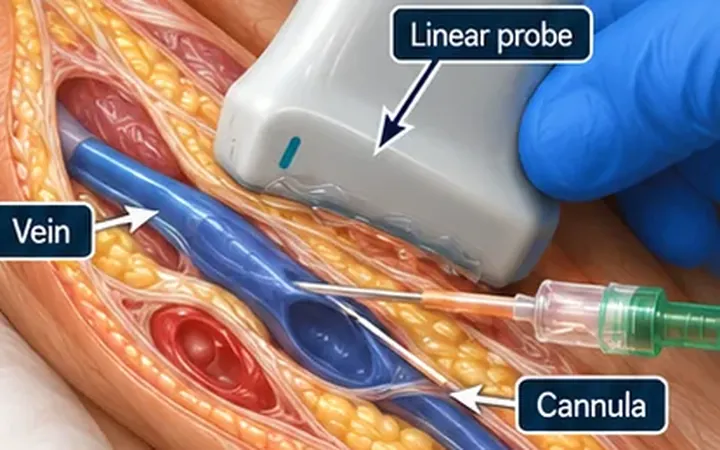
Threading failure: rescue in long axis
Short axis finds the vein and long axis saves the line. When the tip is in the vein but the catheter will not advance, rotate to long axis and look for wall tenting, a valve or a poor angle.
Vascular access
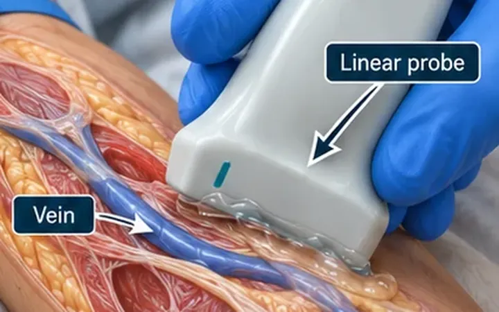
Distension tricks: make the vein come to you
Before you scan, fill the veins. A tourniquet, a dependent arm, warmth and a relaxed squeeze of the hand turn a marginal vein into a usable one.
Vascular access
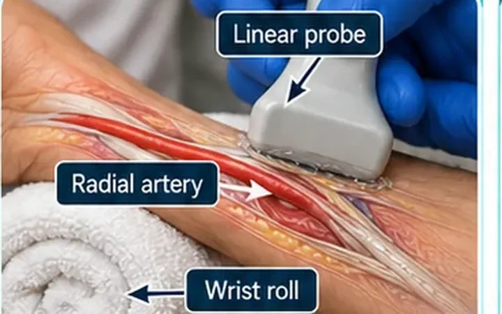
Radial arterial line: trace the pulsation
Pick a straight, superficial stretch of radial artery, steady the wrist and enter at a low angle. If the puncture works but the catheter will not go in, rotate to long axis.
Nothing matches your search.
Try a shorter word, such as "lung" or "abscess", or .
Working towards accreditation?
From learning to sign-off.
Newsletter
Ultrasound skills, in your inbox.
One email a week: a practical scanning skill, new Learn posts and course dates before they go public.