The right upper quadrant view is the most sensitive single view in the FAST scan for free intraperitoneal fluid. Many clinicians stop at Morison's pouch, the space between the liver and the right kidney. Small volumes of fluid often appear first further down, at the inferior tip of the liver and around the lower pole of the kidney.
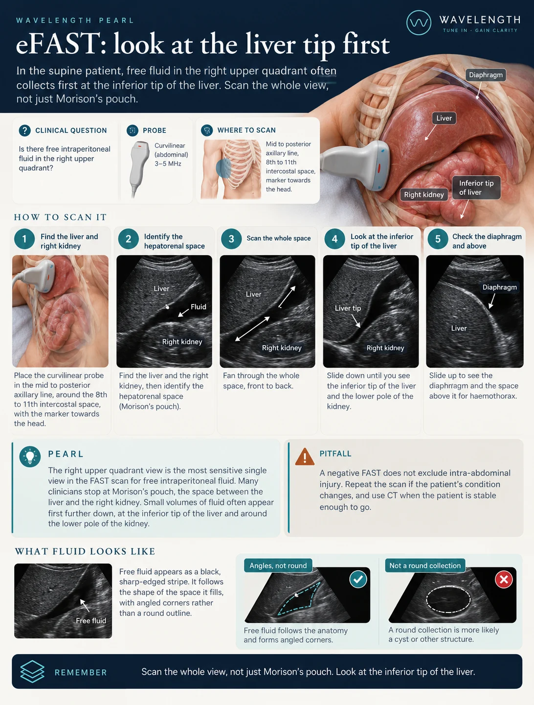
Scan images in the graphic are illustrations, not patient scans.
How to scan it
- Place the curvilinear probe in the mid to posterior axillary line, around the 8th to 11th intercostal space, with the marker towards the head.
- Find the liver and the right kidney, then identify the hepatorenal space.
- Fan through the whole space, front to back.
- Slide down until you see the inferior tip of the liver and the lower pole of the kidney.
- Slide up to see the diaphragm and the space above it for haemothorax.
What fluid looks like
Free fluid appears as a black, sharp-edged stripe. It follows the shape of the space it fills, with angled corners rather than a round outline. A round black structure with a smooth wall is more likely the gallbladder, a cyst, the IVC or fluid-filled bowel.
Why a negative scan does not rule out injury
FAST is specific but misses injuries. A negative scan lowers the chance of a large bleed at that moment. It does not exclude intra-abdominal injury.
- Small volumes. Early in the course of a bleed, the volume of fluid sits below what the scan detects.
- Timing. Fluid builds up over time. A scan in the first minutes after injury underestimates it.
- Patient factors. Obesity, subcutaneous emphysema and dressings all degrade the image.
- Injury patterns. Hollow viscus, diaphragm and retroperitoneal injuries produce little or no free fluid.