Consolidated lung looks like liver on ultrasound: solid, grey and textured. Inside it, bright white dots and lines show air trapped in the airways. These are air bronchograms. One still frame shows they are there. A few seconds of breathing tells you more.
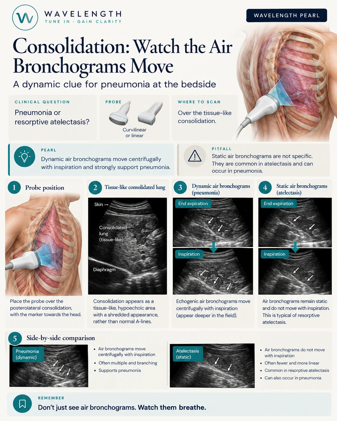
Scan images in the graphic are illustrations, not patient scans.
The scan
- Use the curvilinear probe for the bases and deeper consolidation, or the linear probe for small areas near the pleura.
- Scan the area of concern in both planes, with the marker to the head in long axis.
- Find the tissue-like lung. Its deep edge is often ragged where it meets aerated lung (the shred sign).
- Hold still and watch the bright air bronchograms through two or three breaths.
Dynamic or static
- Dynamic air bronchograms. The bright dots move outwards, away from the centre, as the patient breathes in. Air is still flowing through an open airway. This strongly supports pneumonia.
- Static air bronchograms. The bright dots stay still. The airway is blocked, so air does not move. This is common in resorptive atelectasis, from a mucus plug or an obstructed bronchus, and also seen in some pneumonias.
Other clues
- Fluid bronchograms, black branching tubes inside consolidation, suggest fluid-filled airways behind an obstruction. Colour Doppler shows they carry no flow, unlike vessels.
- A parapneumonic effusion next to consolidation supports infection.
- Compare both sides, and look for B-lines in the surrounding lung.