A red, hot, swollen area raises one question: cellulitis or abscess? Cellulitis needs antibiotics. An abscess needs drainage. Examination often cannot tell them apart. Ultrasound can, in a minute or two.
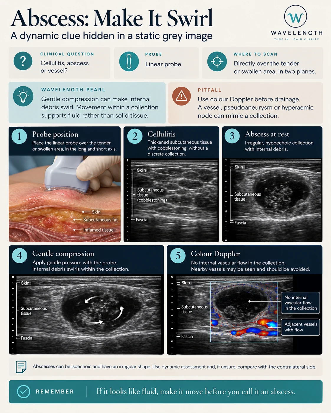
Scan images in the graphic are illustrations, not patient scans.
The scan
- Use the linear probe with plenty of gel, and light pressure on a tender area.
- Start over normal skin, then move over the swelling.
- Scan in two planes, and measure the collection in three dimensions.
- Press gently and release, and watch the contents.
- Switch on colour Doppler over and around the collection.
- If unsure, compare with the same area on the other side.
Reading the view
- Cellulitis. Thickened, bright subcutaneous fat broken up by dark strands of fluid: a cobblestone pattern. No discrete collection.
- Abscess. A dark or mixed collection, often irregular, with brighter debris inside and a brighter rim of tissue. Posterior acoustic enhancement often shows behind it.
- Swirl. Gentle pressure makes the contents move. Movement shows liquid, even when the collection looks grey.
- Colour Doppler. No flow inside an abscess. Flow is often seen around it, in the inflamed rim.
Before drainage
- Mark the deepest point and the best approach.
- Measure the depth from skin to collection.
- Note any vessel nearby, and plan to avoid it.
- In the neck, groin and near joints, think about specialist referral.