A suspected proximal deep vein thrombosis (DVT, a clot in the deep veins of the thigh or behind the knee) is one of the most common reasons to pick up a probe in the emergency department. New scanners often spend their time looking for clot. Fresh clot is often anechoic, black like flowing blood, so you rarely see it. You find it by pressing.
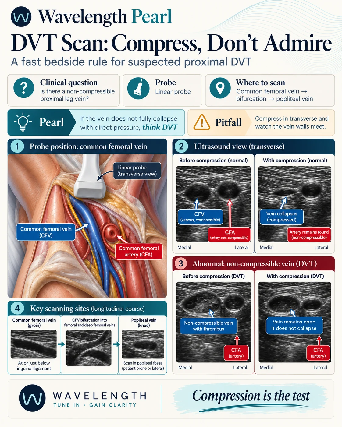
Scan images in the graphic are illustrations, not patient scans.
The scan
- Lie the patient supine with the head of the bed raised, the hip turned out and the knee slightly bent. Raising the head fills the leg veins and makes them easier to see.
- Use the linear probe in transverse, across the leg.
- Start at the groin, at or just below the inguinal ligament. Find the common femoral artery (CFA) and the common femoral vein (CFV), which sits medial to it.
- Press straight down until the vein walls meet. Release, slide the probe 1 to 2 cm down the leg, and press again.
- Keep going past the great saphenous vein junction and the CFV bifurcation, where it divides into the femoral and deep femoral veins.
- Move to the popliteal fossa, with the patient prone or on their side. The popliteal vein lies between the probe and the popliteal artery. Compress it down to where it divides in the upper calf.
The aha
Press until the artery starts to change shape. Then you know you have used enough pressure. If the artery dents and the vein walls meet, the segment is compressible. If the artery dents and the vein stays open, even partly, treat it as clot.
Pitfalls
- A vein which narrows by half and stops is not compressible. Partial compression means non-occlusive clot.
- Groin lymph nodes look round and dark in one still frame. Sweep up and down: a vessel runs on, a node ends.
- Some patients have two femoral veins. Compress both.
- A negative point-of-care scan covers the segments you scanned, not the calf. Follow your local DVT pathway, which usually combines the Wells score, D-dimer and a repeat or formal scan within a week.