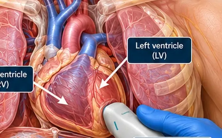
The IVC is easy to find and easy to measure, so it often gets the final word on fluids. Its size and collapse reflect right atrial pressure, which breathing, ventilation, the right heart and the abdomen all change. Use it as one clue among several.
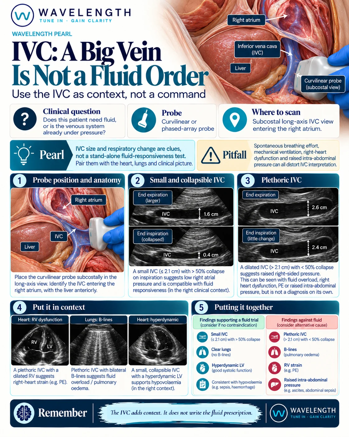
Scan images in the graphic are illustrations, not patient scans.
The scan
- Find the IVC in long axis from the subcostal window with the curvilinear or phased-array probe, and follow it into the right atrium so you know it is not the aorta.
- Measure about 2 cm from the right atrium, just beyond the hepatic vein, at end expiration.
- Watch it with quiet breathing, then with a sniff, and estimate the collapse.
- Scan the heart: right ventricle size, left ventricle function and the pericardium.
- Scan the lungs for B-lines.
Reading the view
- Small and collapsing: 2.1 cm or less, collapsing more than 50% with a sniff, suggests a low right atrial pressure. In a hypotensive patient with a vigorous heart and clear lungs, this supports a cautious fluid trial.
- Large and fixed: over 2.1 cm, collapsing less than 50%, suggests a raised right atrial pressure. Fluid overload, right heart strain, tamponade, tension pneumothorax and raised abdominal pressure all cause it.
- In between: the IVC adds little. Rely on the rest of the assessment.
Putting it together
A plethoric IVC with a dilated right ventricle points to right heart strain. A plethoric IVC with bilateral B-lines points to fluid overload. A small, collapsing IVC with a hyperdynamic left ventricle and clear lungs fits hypovolaemia or vasodilatation. Reassess after every fluid bolus.