Wavelength Pearls
Cardiac and vascular ultrasound.
Cardiac views, the venous system, the aorta and the leg veins.
Focused cardiac ultrasound answers a few questions in a breathless or shocked patient: is the left ventricle squeezing, is the right ventricle under strain, is there fluid around the heart, and how full is the venous system? These pearls take each question in turn, with the view, the measurement and the trap that catches new scanners.
The vascular pearls cover compression ultrasound for proximal DVT, the Baker's cyst which mimics it, and the femoral pseudoaneurysm after a puncture. Each one ends with a short test.
9 pearls
One technique at a time.
Cardiac
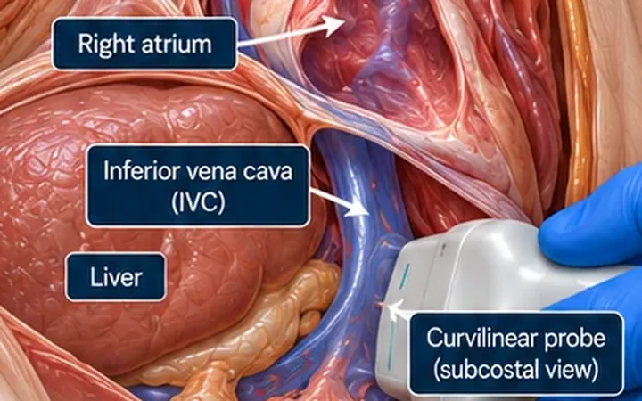
IVC: a big vein is not a fluid order
IVC size and collapse estimate right atrial pressure. They do not tell you whether fluid will help. Read the IVC with the heart, the lungs and the patient.
Cardiac
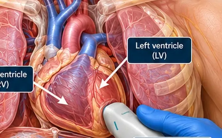
RV strain: three clues beat one ratio
A big right ventricle is a clue. A big right ventricle with a flattened septum, a plethoric IVC and a clot in the leg is a pattern. Build the pattern before you act on it.
Vascular
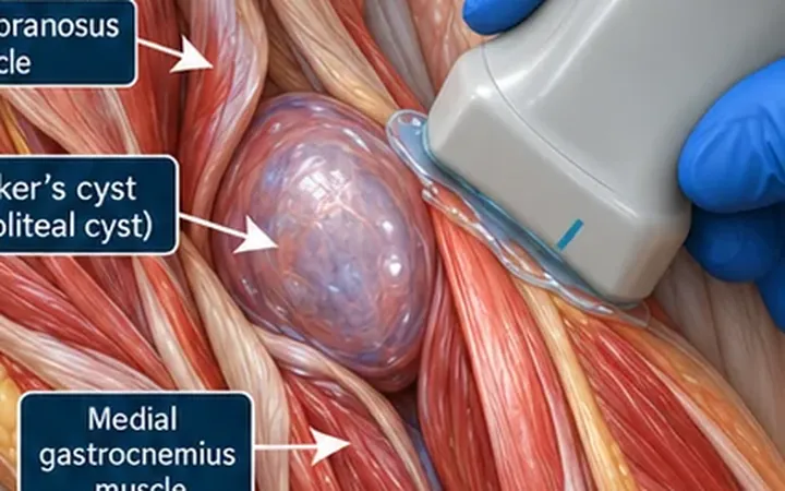
Calf swelling: look behind the knee
A ruptured Baker's cyst mimics a DVT. Find the cyst between semimembranosus and the medial head of gastrocnemius, follow any leaked fluid down the calf, then compress the veins anyway.
Vascular
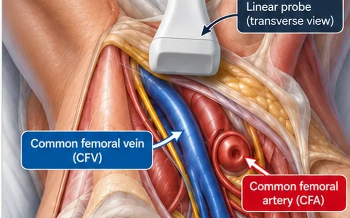
DVT: compress, don't admire
Fresh clot is often black on screen, so a vein full of thrombus looks like a normal vein. The test is whether the walls meet when you press, in transverse, at every site.
Cardiac
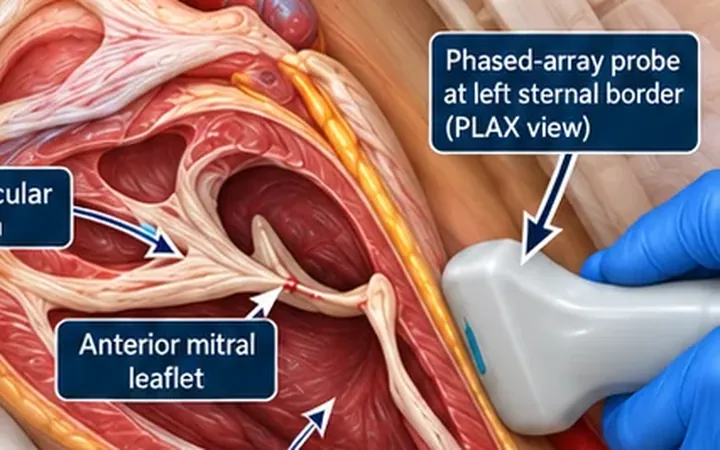
EPSS: when the mitral valve misses the septum
In a strong heart, the anterior mitral leaflet swings open almost to the septum. In a weak heart, it falls short. Measure the gap in the parasternal long-axis view for a quick, objective clue to left ventricular function.
Cardiac
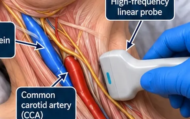
Ultrasound JVP: find the collapse point
The jugular venous pressure is hard to see in a large neck, a busy room or a breathless patient. Follow the internal jugular vein up from the clavicle until it collapses. That point is the top of the venous column.
Cardiac
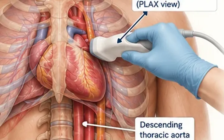
Pericardial or pleural? Find the descending aorta
Black fluid behind the heart in the parasternal long-axis view is either pericardial or pleural. The descending thoracic aorta tells you which. Pericardial fluid runs in front of it. Pleural fluid stays behind it.
Cardiac
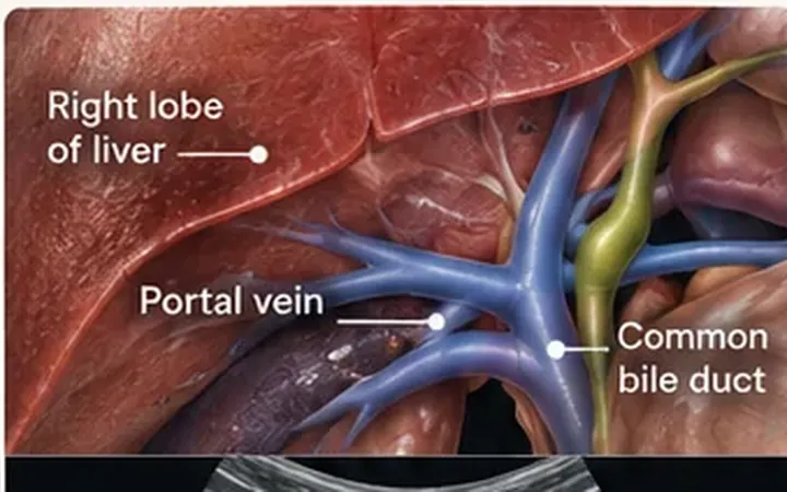
Portal vein pulsatility: a congestion clue
Portal vein flow is normally steady. When pressure backs up from the right heart, it starts to pulse. Read it alongside the IVC, hepatic veins and kidneys, not on its own.
Vascular
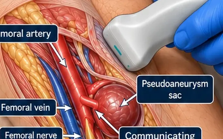
Pseudoaneurysm: find the neck
A pulsatile lump after a femoral puncture is a haematoma, an abscess or a pseudoaneurysm. The swirling sac catches your eye, but the neck makes the diagnosis. Find the channel to the artery and sample it.
Practise it with us
One instructor to every four or five delegates, FAMUS-accredited instructors and RCEM-approved ultrasound supervisors, and long, supervised time on the probe.
Newsletter
Get the next one by email.
One email a week: a practical scanning skill, new Learn posts and course dates before they go public.