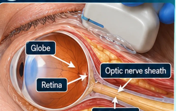
A blow to the eye can tear the fibres that hold the lens, so the lens tilts (subluxation) or falls free (dislocation). Patients notice blurred or double vision in that eye. Marfan syndrome and homocystinuria make it more likely after minor trauma. Ultrasound through the closed lid finds the lens quickly, and the same scan looks for the other causes of sudden loss of vision in the ocular Pearl.
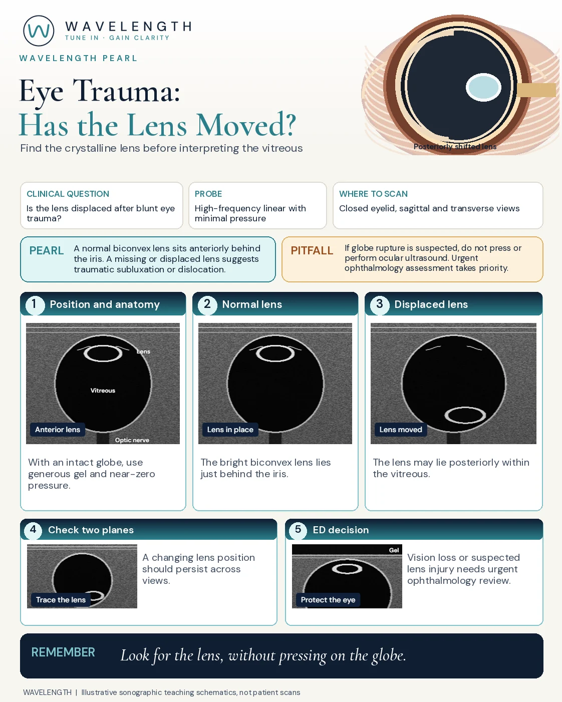
Scan images in the graphic are illustrations, not patient scans.
The scan
- First ask: could the globe be ruptured? If so, stop here.
- Use a high-frequency linear probe with a low-output (ocular) preset if your machine has one.
- Ask the patient to close the eye, cover the lid with plenty of gel and rest your hand on the face so the probe barely touches.
- Scan in transverse and sagittal planes, with the eye looking straight ahead.
- Ask the patient to look side to side, and watch what moves.
Reading the view
- Normal: the lens at the front of the globe, just behind the iris: an oval with bright front and back edges and a dark centre. The vitreous behind is black.
- Posterior dislocation: the lens lies in the vitreous, often on the posterior wall, and is missing from its place behind the iris. It may move as the eye moves.
- Subluxation: the lens is tilted or shifted to one side.
- Anterior dislocation: the lens lies forward in the anterior chamber. This can raise the eye pressure and needs emergency treatment.
Action
Record visual acuity. Any suspected lens injury, or loss of vision, needs urgent ophthalmology review. Shield the eye, give analgesia and an antiemetic, and avoid anything that raises eye pressure.