The optic nerve sheath is continuous with the meninges, so a rise in intracranial pressure can distend it within minutes. The bedside scan takes a few minutes in a patient you are already worried about: headache, vomiting, reduced consciousness or a head injury. Studies report good accuracy, but they use different cut-offs, mostly between about 5 and 6 mm in adults, and the measurement depends heavily on technique.
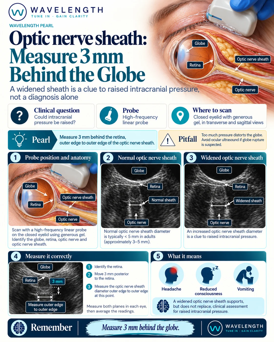
Scan images in the graphic are illustrations, not patient scans.
The scan
- Do not scan if you suspect an open globe.
- Select the ophthalmic or ocular preset, which keeps the power within the limits set for the eye.
- Ask the patient to close the eye and look straight ahead. Cover the eyelid with a thick layer of gel and rest your hand on the face, so the probe floats with no pressure.
- Find the dark band of the optic nerve running straight back from the globe, centred on the screen.
- Measure 3 mm back from the retina along the nerve, then measure across the sheath at that depth, outer edge to outer edge.
- Repeat in the other plane and in the other eye. Average the readings.
Reading the view
- Normal: a sheath up to about 5 mm across in adults; smaller in children.
- Wide: above about 5 to 6 mm in adults. A clue to raised intracranial pressure.
- Supporting signs: a bulging optic disc into the globe (papilloedema) adds weight.
Action
Do not delay CT, neurosurgical advice or treatment to scan the eye. Use a wide sheath to escalate faster, and record both eyes and both planes so the next measurement can be compared.