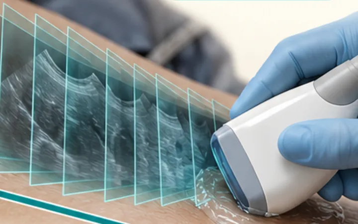
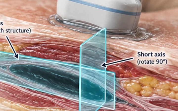
The machine assumes every echo travelled straight out and straight back. At a strong curved reflector such as the diaphragm, with air-filled lung behind it, some sound takes a longer path. The machine places those late echoes deeper than they belong, and paints a copy of the liver or spleen above the diaphragm.
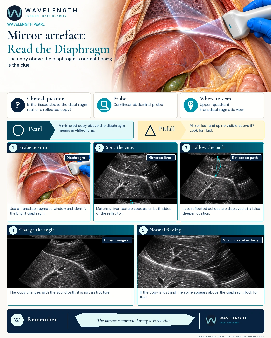
Scan images in the graphic are fabricated educational illustrations, not patient scans.
Recognise it
- Find the bright curved diaphragm from the right or left upper quadrant.
- Compare the tissue on each side of it.
- A mirror image repeats the organ below, with the same texture and often the same vessels.
- Change the probe angle or the window. The copy changes with the sound path, because it is not a structure.
What it tells you
- Mirror image present: the lung above the diaphragm is full of air. In the eFAST and lung scan this is the normal finding.
- Mirror image lost, with the spine visible above the diaphragm: the "spine sign". Fluid in the chest lets sound through, so you see the vertebrae continue upwards. Think pleural effusion or haemothorax.
- Tissue-like lung above the diaphragm, with moving bright air bronchograms: consolidation, not a mirror. Real lung keeps chest anatomy; a mirror copies the abdomen.
Action
Move, angle and compare. Use a second view before you record an unexpected finding above the diaphragm, and read the result with the pleural effusion pearl's spine sign.