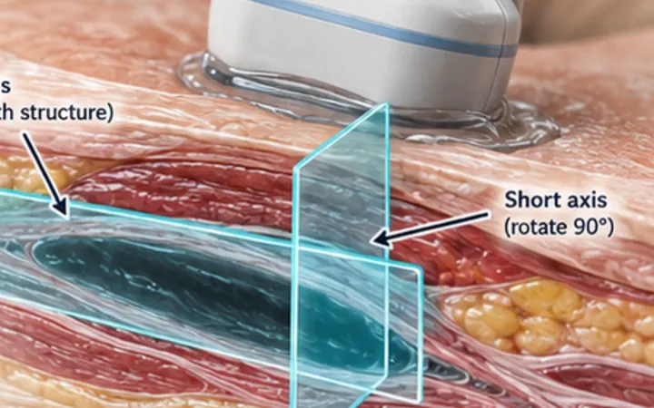
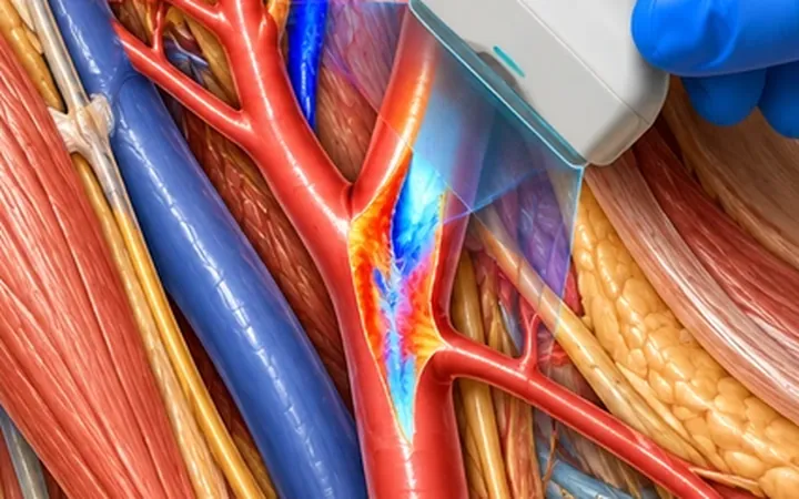
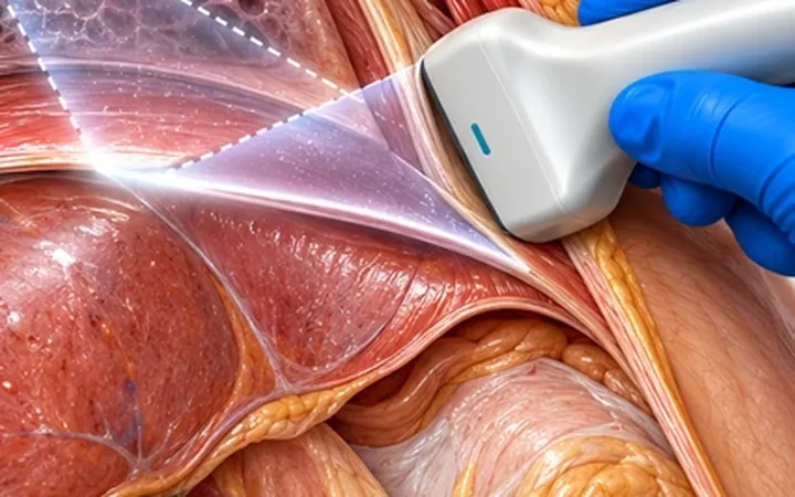
An ultrasound image is a slice about a millimetre or two thick. A small collection, a stone, a fluid pocket or a foreign body can sit just beside it. A good scan is a survey through the whole target, and the saved image is the record at the end of it, not the scan itself.
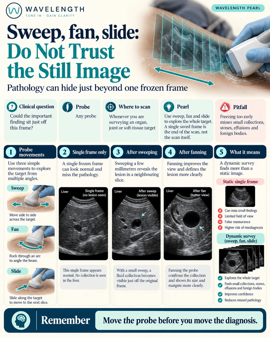
Scan images in the graphic are illustrations, not patient scans.
The movements
- Sweep: move the probe sideways across the target, keeping the same angle, to see neighbouring slices.
- Fan: keep the probe still on the skin and tilt it through an arc, so the beam passes through a volume beneath one point. Useful between ribs or under a narrow window.
- Slide: move the probe along the long axis of a structure to follow it, such as a vessel, a tendon or the bowel.
- Add rotation to turn between long and short axis, as in the two planes pearl.
Using them
- Survey first, from one edge of the target to the other, before freezing anything.
- Move slowly: about a second for each centimetre lets the eye catch small findings.
- When you find something, sweep through it to define its edges, then record it in two planes.
Action
Make the survey a habit for every application. When you record a scan, note the views and the movement used, so the next person can repeat it.