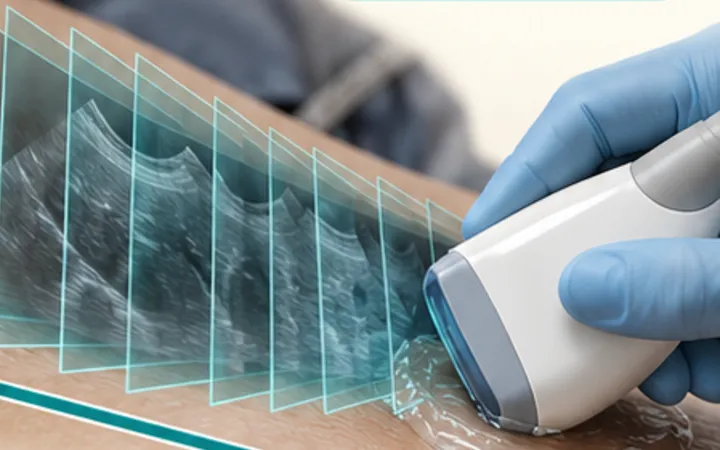
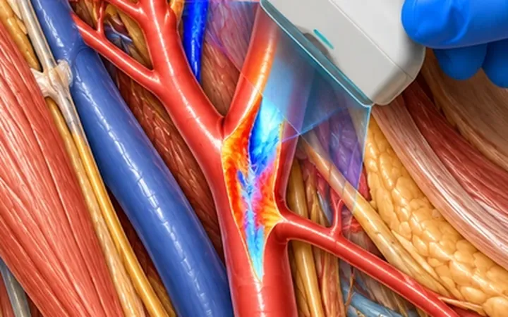
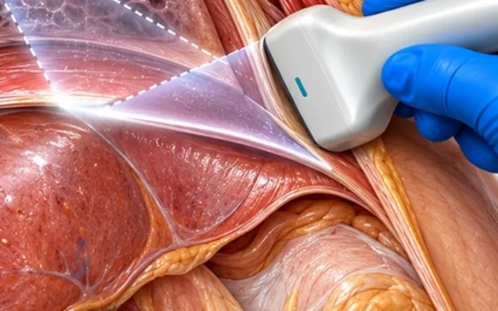
Every ultrasound image is a slice through a three-dimensional body. A round structure can be a tube cut across; a dark gap in a tendon can be anisotropy; a fluid pocket can be a fold of bowel. Turning the probe through 90 degrees answers most of these questions in seconds.
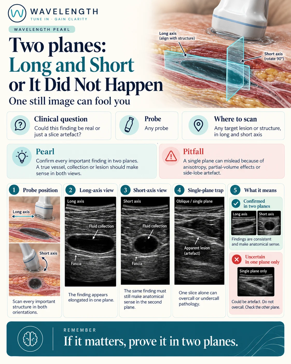
Scan images in the graphic are illustrations, not patient scans.
The scan
- Find the structure and centre it on the screen.
- Line the probe up with its long axis and record that view.
- Keep it centred and rotate the probe 90 degrees for the short axis.
- Sweep through it in both planes, as in the sweep, fan, slide pearl.
- Measure in both planes when size matters.
Reading the view
- Vessel: a tube in long axis, a circle in short axis, continuous as you sweep.
- Collection: a defined pocket in both planes, often oval in one and round in the other.
- Artefact or partial volume: looks convincing in one plane and disappears or changes shape in the other.
- Anisotropy: a dark patch in a tendon that changes with probe angle; see the anisotropy pearl.
Action
Record important findings in both planes, with measurements in both when size matters. Before any needle procedure, check the target and the path in both planes.