Cervical artery dissection is a tear in the arterial wall. Blood tracks into the wall, narrows the lumen and seeds clots that travel to the brain. It causes up to a fifth of strokes in people under 45, often after minor neck trauma or sudden neck movement. The clues are one-sided neck pain or headache, a partial Horner syndrome (a small pupil and a drooping lid), pulsatile tinnitus, and a transient or persisting neurological deficit.
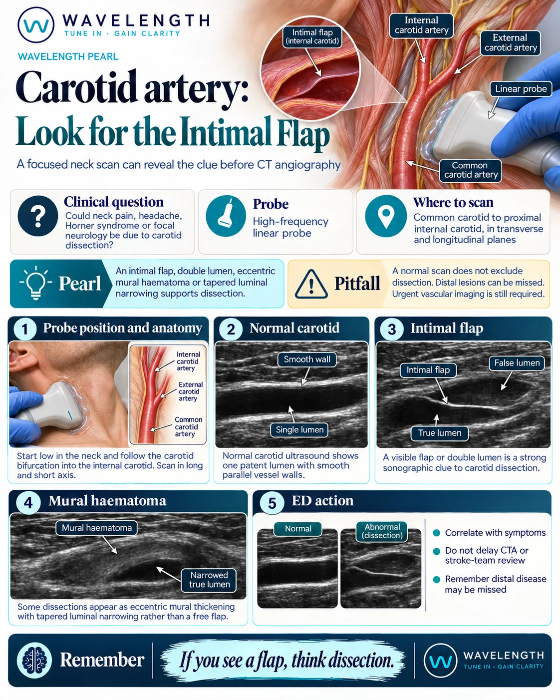
Scan images in the graphic are illustrations, not patient scans.
The scan
- Use a high-frequency linear probe with the head turned slightly away.
- Start low on the common carotid artery in transverse and follow it up to the bifurcation.
- Follow the internal carotid artery as far up towards the jaw as it can be seen, in transverse and long axis.
- Add colour and spectral Doppler in the common and internal carotid arteries, and compare with the other side.
- If you can, look at the vertebral artery between the transverse processes as well.
Reading the view
- Normal: one lumen with smooth, parallel walls and colour filling the whole lumen.
- Intimal flap: a thin bright line inside the lumen that moves with each pulse, with flow on both sides: a true and a false lumen.
- Mural haematoma: a crescent of dark, eccentric wall thickening that narrows the lumen and tapers over a length of artery, often without a free flap.
- Indirect signs: a high-resistance waveform with little or no diastolic flow, or very low flow, in the internal carotid, which suggests narrowing or occlusion higher up.
Action
Suspected dissection with neurology goes on the stroke pathway at once: the scan must not delay CT angiography, the stroke team or thrombolysis and thrombectomy decisions. Without neurology, arrange urgent CT or MR angiography. Antiplatelet or anticoagulant treatment is decided by the stroke team.