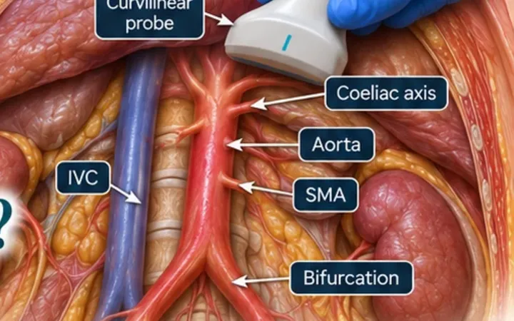
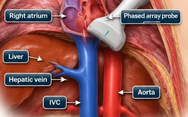
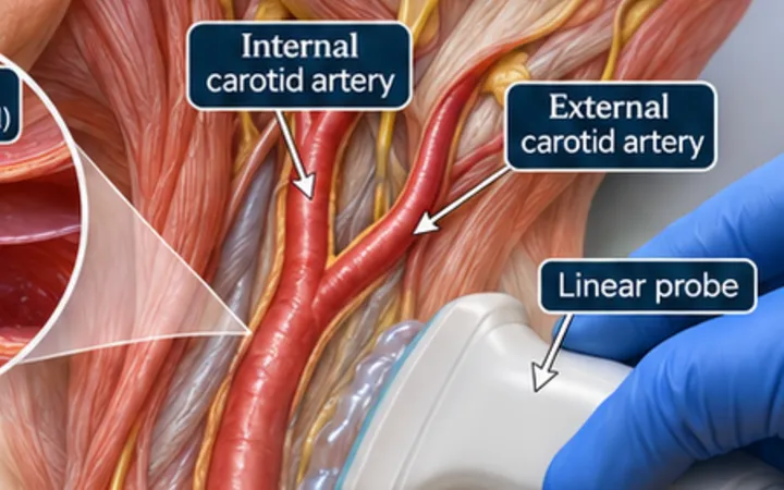
The subcostal view is the window you need most in the sickest patients: during cardiac arrest, in the ventilated patient, and when the chest is hard to scan. It is also the view new scanners find hardest. The reason is nearly always the angle of the probe, not the patient.
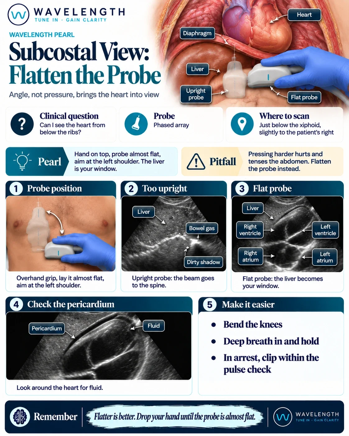
Scan images in the graphic are illustrations, not patient scans.
Before
Holding the probe like a pen, pointing straight into the abdomen, sends the beam back towards the spine. You see liver near the probe, then bowel gas with a grey, dirty shadow beneath it. The heart sits above and behind the ribs, out of the beam.
The move
- Ask the patient to lie flat and bend the knees. This relaxes the abdominal wall.
- Use the phased array probe with the marker to the patient's left.
- Change your grip: put your hand on top of the probe, palm down, so you hold it from above.
- Place the probe just below the xiphoid, slightly to the patient's right, so the liver becomes your window.
- Lay the probe almost flat against the skin and aim towards the left shoulder.
- Increase the depth, then ask the patient to take a deep breath in and hold it. This brings the heart down towards the probe.
After
The liver now fills the near field, with the heart behind it. The right ventricle sits nearest the probe, against the liver, with the left ventricle beyond it. The atria lie to the left of the screen and the apex points to the right. The pericardium is the bright line around the heart, and this is where you look for a pericardial effusion.