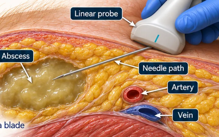
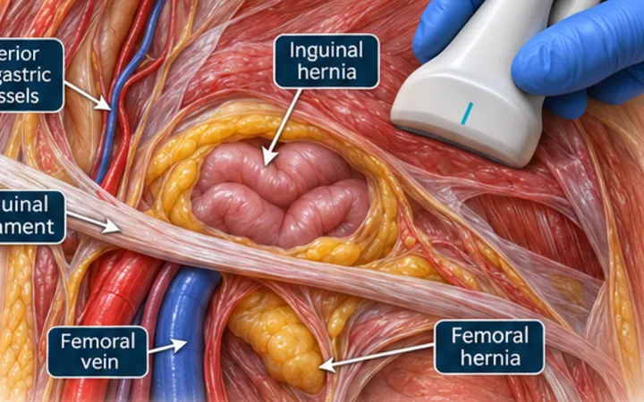
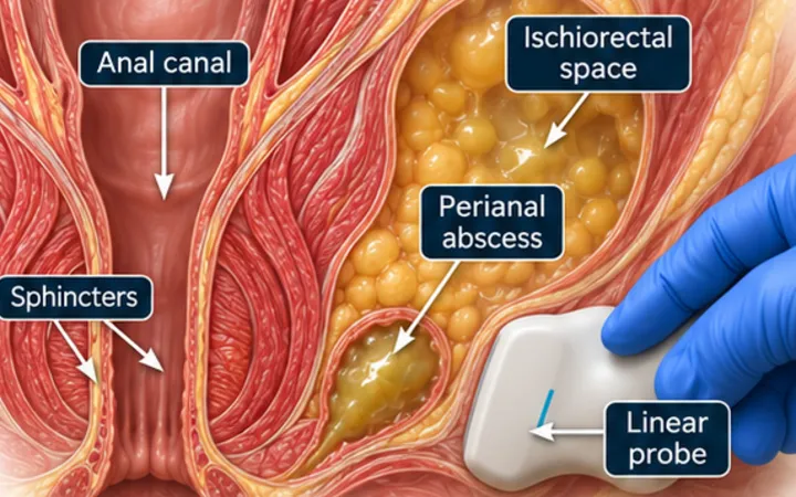
Mastitis is common in breastfeeding women and usually settles with continued feeding or expressing, and antibiotics if needed. A minority go on to form an abscess, which often does not settle on antibiotics alone. Non-lactational abscesses happen too, particularly in smokers, and tend to recur. Ultrasound shows whether there is a pocket to treat, and repeated needle aspiration has replaced surgical incision as the usual first step for many of them.
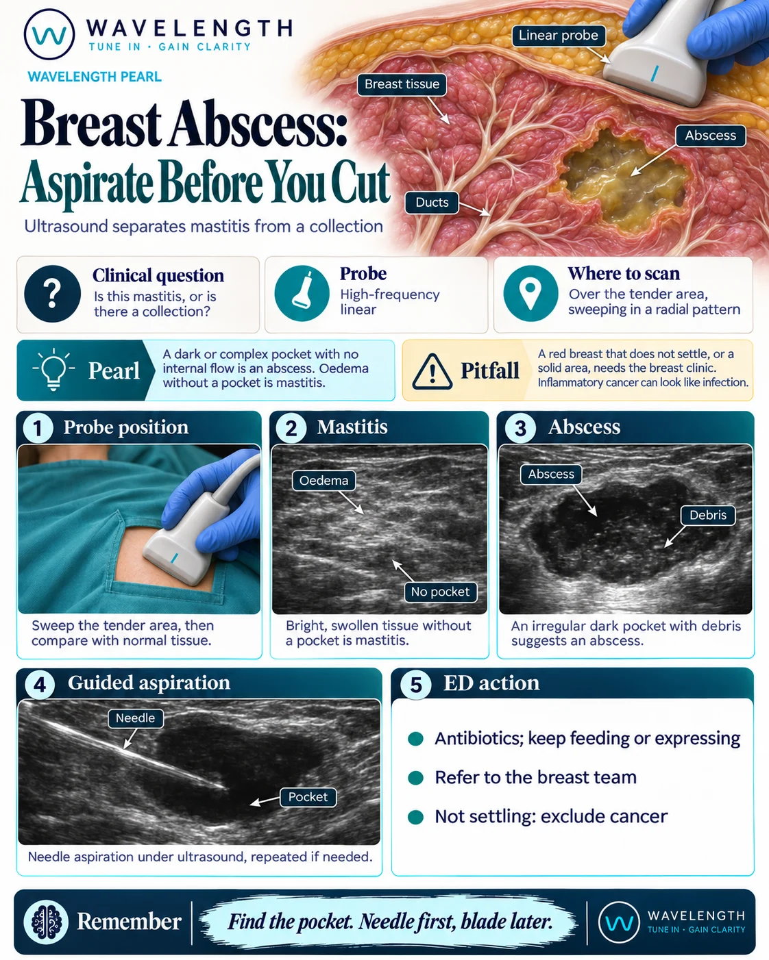
Scan images in the graphic are illustrations, not patient scans.
The scan
- Explain the scan, keep the patient covered, and offer a chaperone.
- Use a high-frequency linear probe with plenty of gel and a light touch.
- Sweep over the tender area in a radial pattern, spreading out from the centre.
- Compare it with normal tissue nearby or with the other breast.
- Add colour Doppler, press gently, and measure any pocket.
Reading the view
- Mastitis: bright, swollen tissue with blurred layers and no pocket.
- Abscess: an irregular dark or complex pocket with debris, no flow inside it, and often flow around it.
- Solid area: a mass that does not behave like fluid needs the breast clinic.
Action
Give antibiotics according to local guidance, and encourage the patient to keep feeding or expressing. Refer to the breast team, who usually offer needle aspiration under ultrasound, repeated if needed. Incision is usually kept for large collections, thin or failing skin, or when aspiration has failed. Sepsis needs admission. If the problem does not settle, arrange assessment to exclude cancer.