Wavelength Pearls
Soft tissue and musculoskeletal ultrasound.
Collections, foreign bodies, tendons and joint effusions.
Soft tissue and musculoskeletal ultrasound settles common emergency department questions in minutes: abscess or cellulitis, a retained foreign body, a torn tendon, fluid in a joint, or a dislocated shoulder before and after reduction.
These pearls cover abscess and necrotising fasciitis, foreign bodies, hernias, Achilles, quadriceps and distal biceps tears, tendon anisotropy, and effusions of the hip, knee and shoulder, each with a short test.
13 pearls
One technique at a time.
Soft tissue
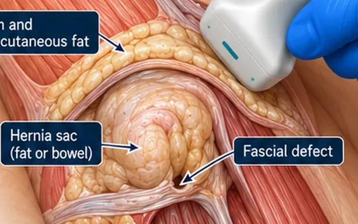
Abdominal wall hernia: stand them up and make it cough
A small hernia hides when the patient lies still. Scan the lump at rest, then during a cough, a strain or standing. Show the defect, what passes through it, and whether it goes back.
MSK
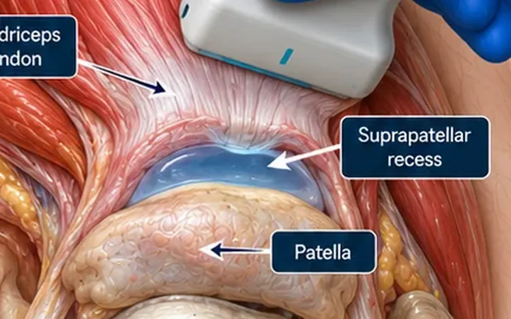
Hot knee: find the suprapatellar recess
Knee fluid collects in the suprapatellar recess, deep to the quadriceps tendon. Find it, squeeze fluid into it, and use it to guide aspiration. Ultrasound finds fluid. It does not tell you if it is infected.
MSK
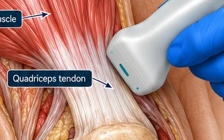
Quadriceps tendon: follow it to the patella
A fall, a giving-way knee and a patient who cannot straight-leg raise. Follow the quadriceps tendon down to the top of the patella and look for a gap. Bend the knee gently, and a true tear opens.
MSK
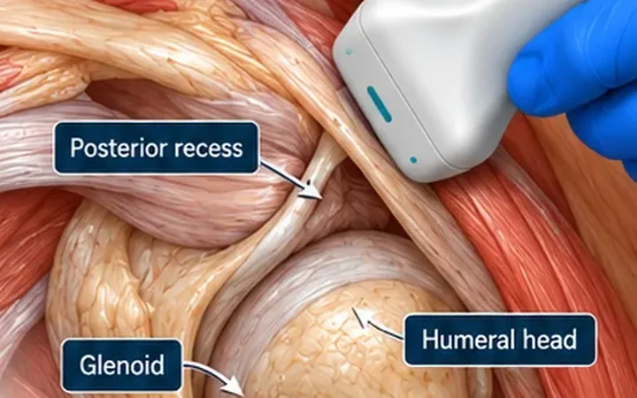
Shoulder effusion: start in the posterior recess
Glenohumeral fluid shows best behind the joint. From the posterior view, look for a dark space lifting the capsule off the humeral head, and compare with the other shoulder.
Soft tissue
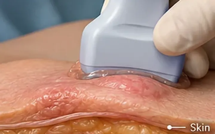
Abscess: make it swirl
An abscess can look grey and solid on a still image. Press gently with the probe. If the contents swirl, it is fluid. Then check colour Doppler before anyone reaches for a needle.
MSK
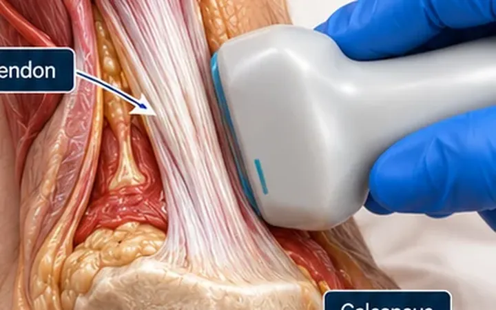
Achilles rupture: scan long, squeeze the calf
A still image of a torn Achilles tendon can look confusing. Scan in long axis, then squeeze the calf or move the ankle while you watch. If the movement stops at a gap, the tendon has ruptured.
MSK
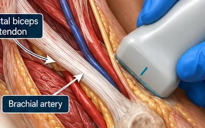
Distal biceps rupture: follow the tendon to the tuberosity
A pop at the elbow while lifting, then weakness and bruising. The tendon can look intact in the elbow crease when it has torn from the bone. Follow it all the way to the radial tuberosity. An empty insertion is the clue.
Soft tissue
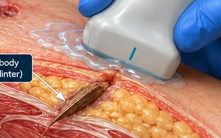
Foreign body: bright, then read the shadow
X-ray misses wood, plastic and thorns. Ultrasound sees every foreign body as a bright structure, and what lies behind it hints at the material. Find it, map it, then cut.
MSK
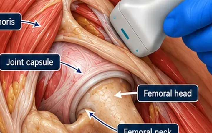
Hip effusion: compare the capsule distance
A limping child or a hot, painful hip raises the question of fluid in the joint. Align the probe with the femoral neck, measure the capsule-to-neck distance, then scan the other hip. Asymmetry is the strongest sign.
Soft tissue
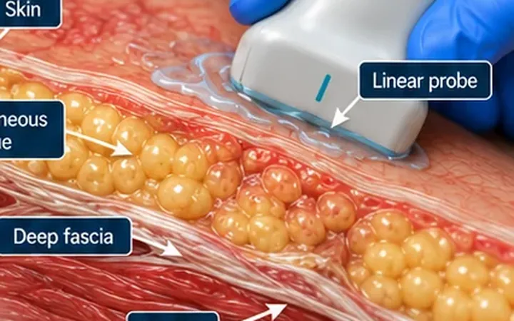
Necrotising fasciitis: look below the cobblestones
Cobblestoning fits cellulitis. Fluid along the deep fascia and gas in the soft tissue raise the alarm for necrotising fasciitis. Use the scan to escalate faster, never to reassure.
Soft tissue
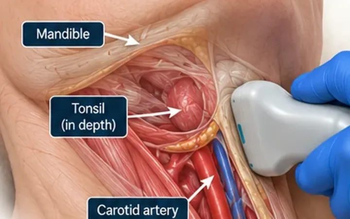
Peritonsillar abscess: scan from the neck
Trismus and gagging make an intraoral look difficult. A linear probe under the angle of the jaw shows whether there is a collection to drain, and where the carotid artery is before anyone reaches for a needle.
MSK
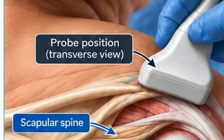
Shoulder: one posterior view, before and after reduction
Put the probe below the scapular spine and the glenoid and humeral head sit side by side. Where the head lies in relation to the glenoid tells you if the shoulder is out, which way, and if it has gone back in.
MSK
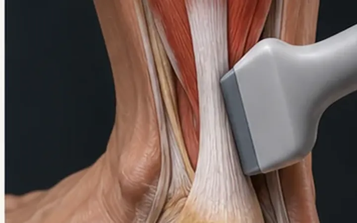
Tendon tear or anisotropy? Heel-toe before you call it
A normal tendon turns dark when the beam hits it at an angle. Rock the probe until the fibres brighten. If the dark area stays, think tear.
Practise it with us
One instructor to every four or five delegates, FAMUS-accredited instructors and RCEM-approved ultrasound supervisors, and long, supervised time on the probe.
Newsletter
Get the next one by email.
One email a week: a practical scanning skill, new Learn posts and course dates before they go public.