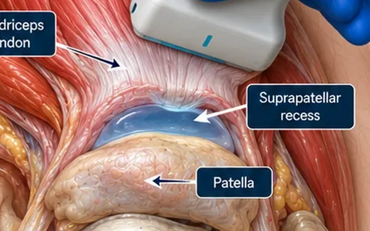
Quadriceps tendon rupture is often missed. Swelling hides the defect, the X-ray looks near normal, and a partial straight-leg raise reassures. Ultrasound shows the tendon directly, and a few seconds of movement settle most doubts.
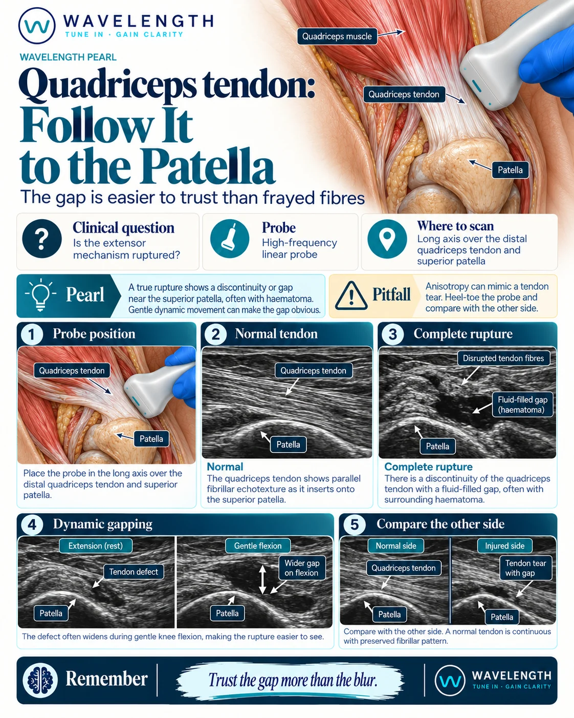
Scan images in the graphic are illustrations, not patient scans.
The scan
- Lie the patient supine with the knee straight or slightly bent over a pillow.
- Place the linear probe in long axis above the patella, marker to the head.
- Find the bright, fibrillar quadriceps tendon and follow it down to where it inserts on the top of the patella.
- Sweep from medial to lateral across the whole width of the tendon.
- Gently bend the knee a little, if pain allows, and watch the insertion.
- Scan the other knee with the same settings.
Reading the view
- Normal: parallel bright fibres run in a continuous band onto the superior patella.
- Complete rupture: a gap in the tendon, usually within 2 cm of the patella, filled with dark fluid or haematoma. The torn ends look wavy and retracted, and the gap widens as the knee bends.
- Partial tear: some fibres are lost, but part of the tendon stays continuous and the gap does not open through the full thickness.
What to do next
Suspected complete rupture needs urgent orthopaedic referral, because early repair gives the best results. Splint the knee straight. Check the X-ray for a low-lying patella or a bony avulsion. Also look at the patellar tendon below the patella, which tears in younger patients.