A tender lump in the abdominal wall, or by a scar, raises the question of a hernia and whether it is stuck. A still image in a supine patient often shows nothing. Movement shows the hernia.
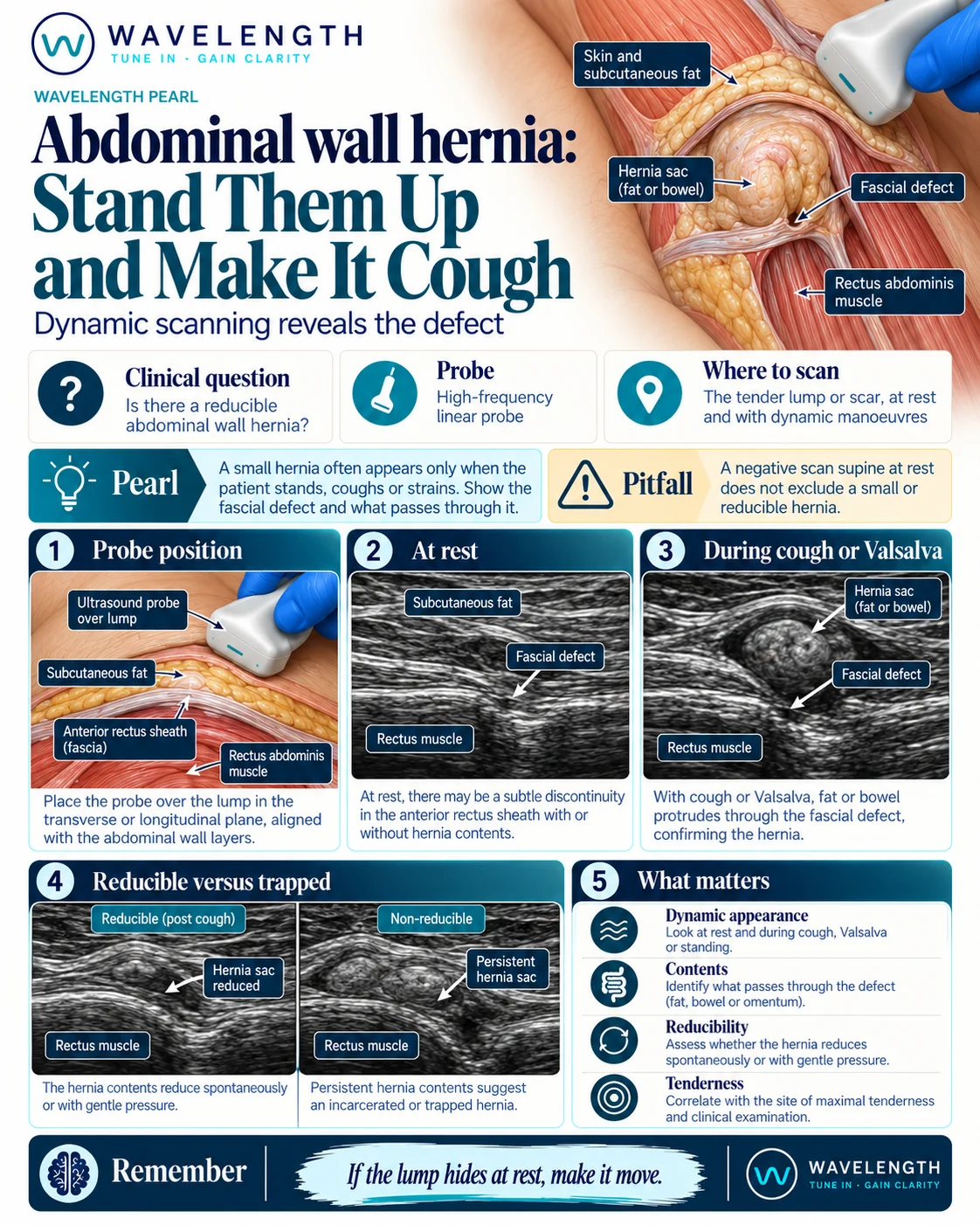
Scan images in the graphic are illustrations, not patient scans.
The scan
- Use the linear probe, or the curvilinear probe in a larger patient.
- Place the probe over the lump or the most tender point, across the abdominal wall layers.
- Identify skin, fat, the anterior rectus sheath and the muscle, then follow the fascia looking for a break.
- Ask the patient to cough or bear down, and watch the defect.
- If the scan is still negative, stand the patient up and repeat.
- Press gently with the probe and watch whether the contents go back.
Reading the view
- Defect: a break in the bright fascial line.
- Contents: fat looks bright and compressible. Bowel shows a layered wall, fluid or gas, and may move.
- Reducible: contents slide back through the defect at rest or with gentle pressure.
- Incarcerated: contents stay in the sac and will not reduce.
- Concern for strangulation: a non-reducible sac with thick-walled bowel, fluid in the sac, no peristalsis, or no flow in the wall on colour Doppler, together with pain and tenderness.
What to do next
A painful, non-reducible hernia, especially with vomiting or signs of obstruction, needs urgent surgical review. A reducible hernia with no red flags can go through the routine surgical pathway.