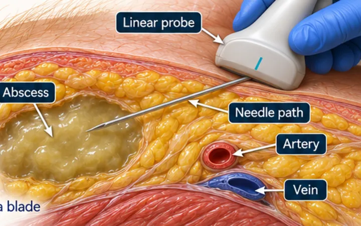
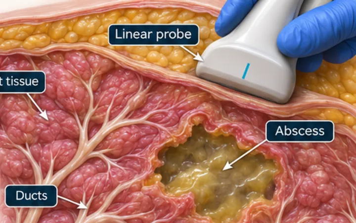
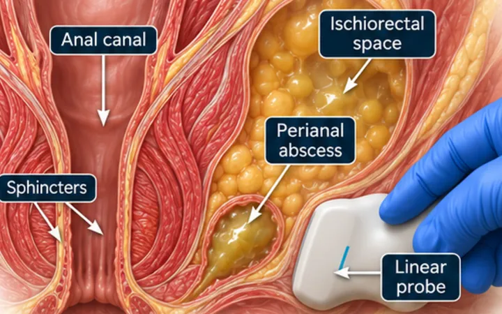
A groin lump may be a hernia, a lymph node, a vein, an artery or an abscess, and the treatment for each is very different. Femoral hernias matter most. They are less common than inguinal hernias, are more common in older women, and carry a high risk of strangulation. The inguinal ligament and the femoral vein are the two landmarks that sort them.
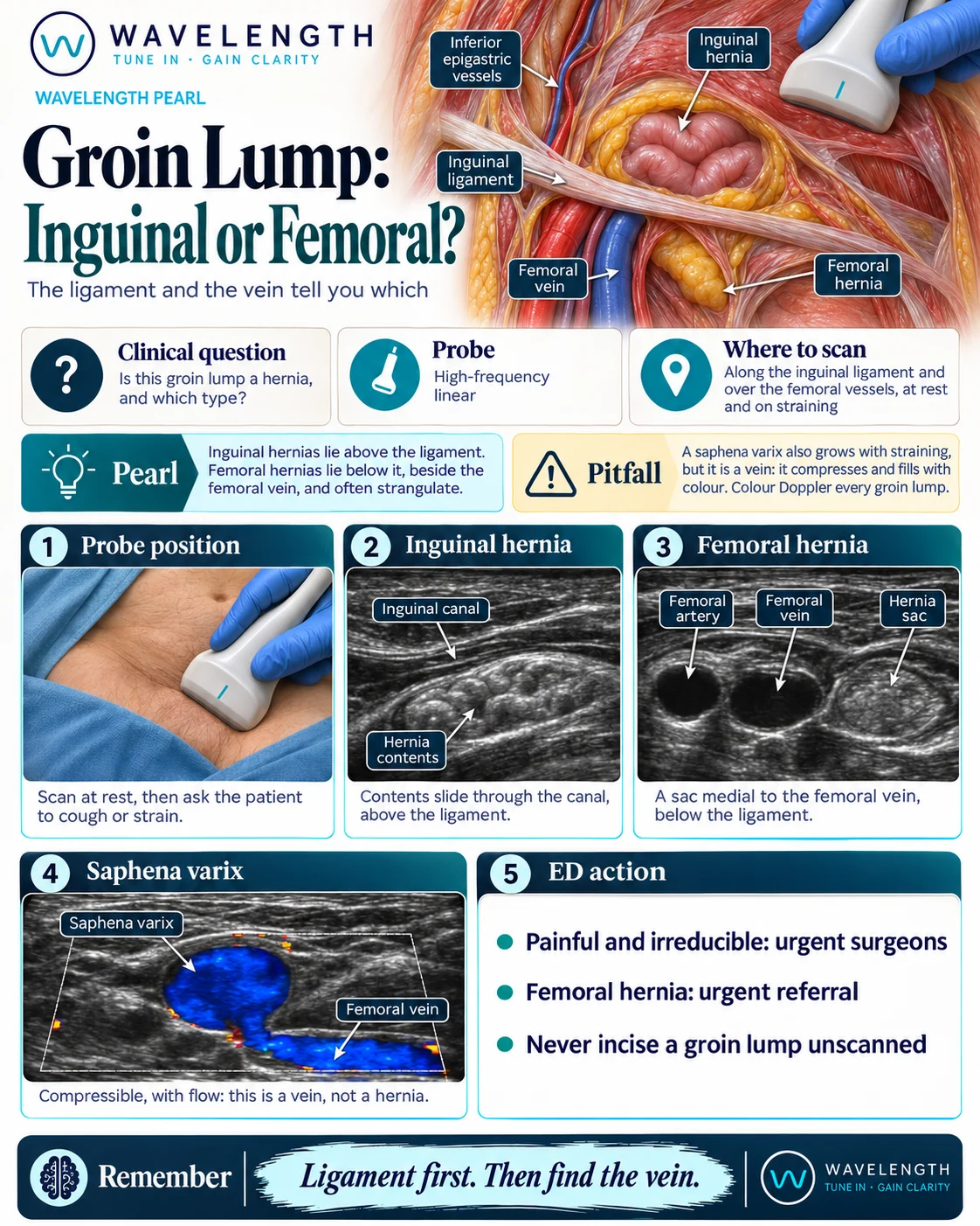
Scan images in the graphic are illustrations, not patient scans.
The scan
- Use a high-frequency linear probe, with the patient lying flat. Stand them up if the first scan shows nothing.
- Find the femoral artery and vein in transverse, below the inguinal ligament.
- Scan along the inguinal ligament and the inguinal canal above it.
- Scan the lump at rest, and then ask the patient to cough or strain.
- Add colour Doppler to every groin lump, and press gently to see whether it compresses.
Reading the view
- Inguinal hernia: contents slide through the canal, above the ligament.
- Femoral hernia: a sac below the ligament and medial to the femoral vein, which may narrow the vein on straining.
- Saphena varix: a round black vessel joining the femoral vein. It compresses and fills with colour.
- Pseudoaneurysm: a pulsatile sac with swirling flow and a neck to an artery.
- Lymph node: an oval with a bright centre, which does not change when the patient strains.
Action
A painful, irreducible lump needs urgent surgical review. A femoral hernia needs urgent referral, even when it is not painful, because it often strangulates. A reducible inguinal hernia with no red flags can go through the routine surgical pathway, with advice about pain, vomiting and a lump that will not go back. Never incise a groin lump that has not been scanned.