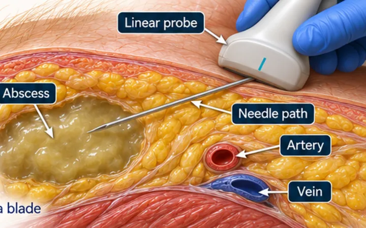
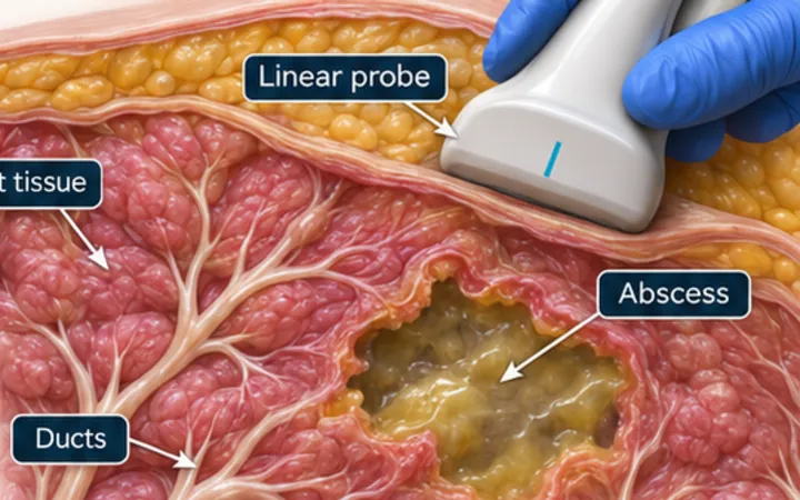
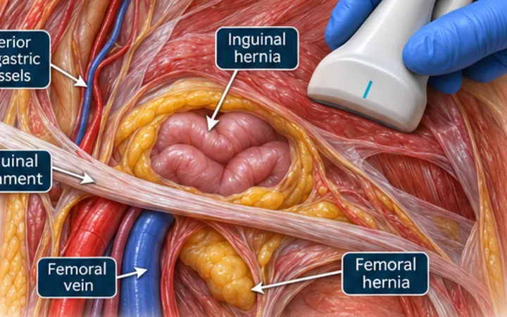
An incarcerated hernia is stuck. A strangulated hernia has lost its blood supply, and the bowel inside it is dying. Examination cannot always separate them, and the delay matters. Ultrasound can show what is in the sac and what is happening upstream, but it supports the surgical decision and does not replace it.
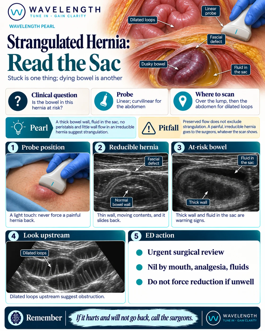
Scan images in the graphic are illustrations, not patient scans.
The scan
- Use a linear probe over the lump, with a light touch. Use the curvilinear probe in a larger patient.
- Find the break in the bright fascial line and what passes through it.
- Decide whether the contents are fat or bowel.
- Watch the bowel for peristalsis, measure the wall, and look for fluid in the sac.
- Add colour Doppler with a low scale, and compare the wall with a normal loop.
- Change to the curvilinear probe and look in the abdomen for dilated loops.
Reading the view
- Reducible hernia: a thin-walled loop with moving contents, which slides back.
- Incarcerated: the contents stay in the sac and do not reduce.
- Bowel at risk: a thick wall, fluid in the sac, no peristalsis and little or no flow in the wall.
- Upstream: dilated, fluid-filled loops with visible folds suggest obstruction.
Action
Arrange urgent surgical review. Keep the patient nil by mouth, and give analgesia and intravenous fluids. Send bloods including a lactate, and treat sepsis if it is present. Do not force reduction in a patient who is unwell, or where the scan suggests bowel at risk, because that can return dead bowel to the abdomen.