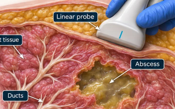
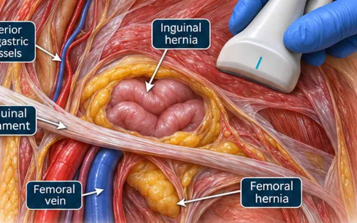
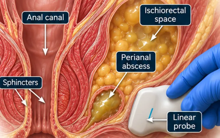
Once you have shown that a lump is a collection, the next question is whether it is safe to drain it here, and by which route. A minute with the probe gives you the depth, the size and the structures to avoid. It also stops the worst error of all: putting a needle into a vessel.
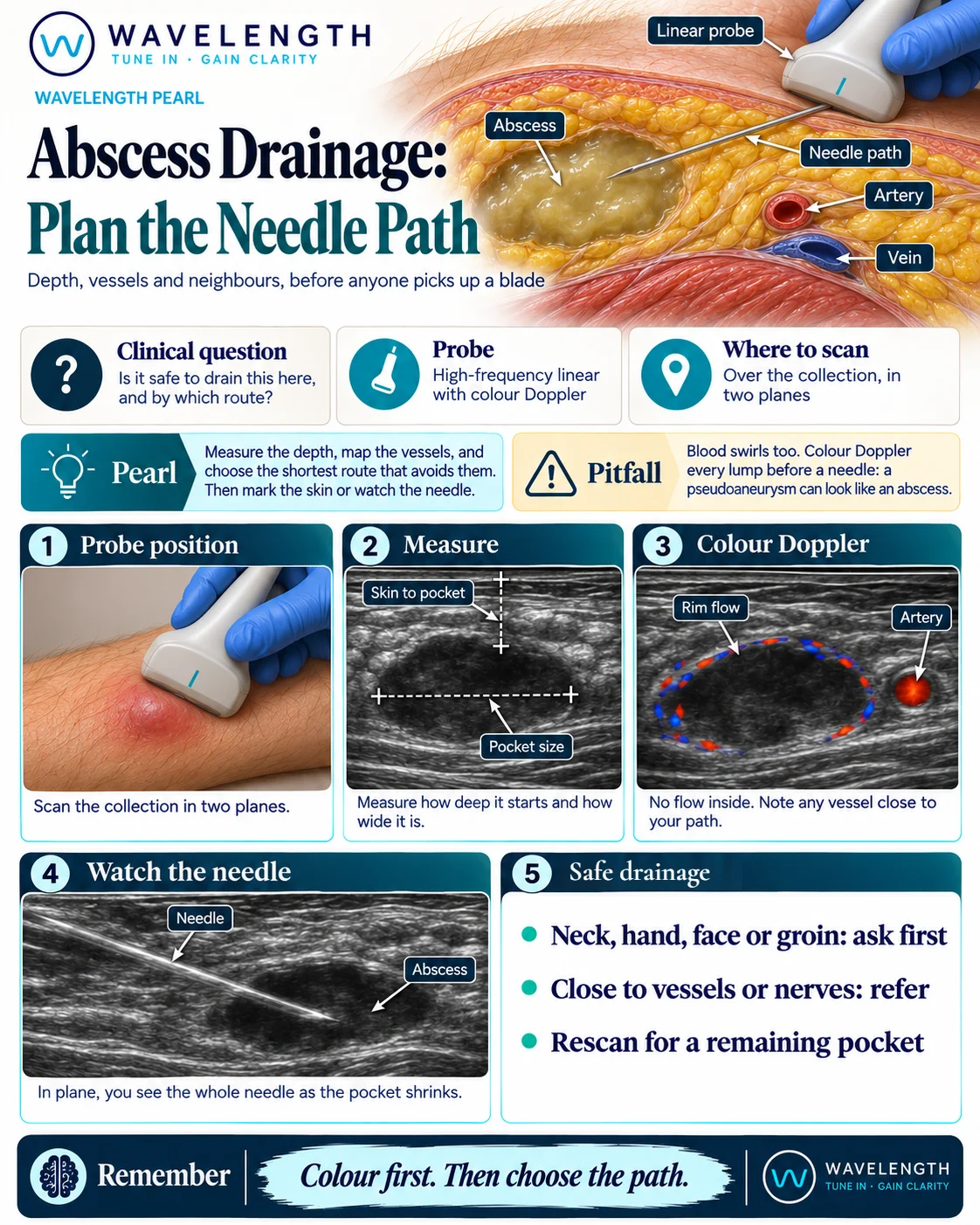
Scan images in the graphic are illustrations, not patient scans.
The scan
- Use a high-frequency linear probe over the collection, in two planes.
- Switch on colour Doppler, with the scale set low. Look inside the lump and around it.
- Measure from the skin to the pocket, and across the pocket.
- Find any artery, vein or nerve close to your path, and anything deep to the pocket.
- Choose the shortest route that avoids them. Mark the skin, or watch the needle under real-time guidance.
- Rescan at the end for a remaining pocket.
Reading the view
- Safe target: a dark or complex pocket with no flow inside it, and sometimes flow in its rim.
- Path: the shortest route to the largest part of the pocket, with no vessel in the way.
- Marked skin: quick, but the mark moves if the patient or the skin moves after the scan.
- In plane: you see the whole needle, and you watch the pocket shrink as you aspirate.
Action
Ask for help before draining a lump in the neck, hand, face or groin, and refer a collection close to vessels or nerves. Any flow inside the lump, or a connection to an artery, means no needle. Suspected necrotising infection needs surgeons, not a local drainage. Record the size, the depth, the Doppler findings and what remained at the end.