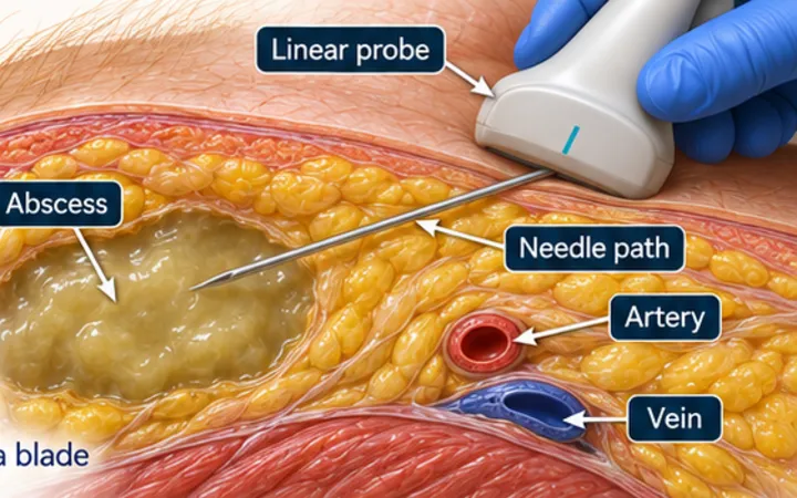
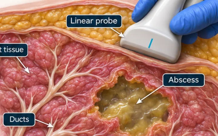
Most anorectal abscesses begin in an infected anal gland and spread through the planes around the sphincters. They may stay close to the skin, or track into the ischiorectal fossa, between the sphincters or above the levator muscle. The size of the lump on the skin tells you little about what lies beneath it, so a scan can help to plan the next step, if the patient can tolerate it.
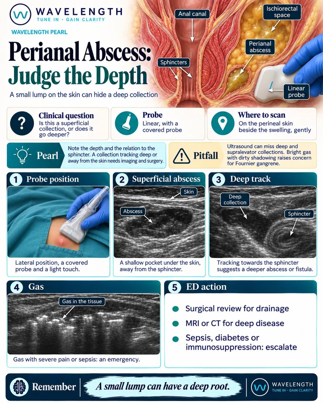
Scan images in the graphic are illustrations, not patient scans.
The scan
- Explain the scan and give analgesia. Lay the patient on their side, with a chaperone and drapes.
- Use a linear probe in a cover, on the perineal skin beside the swelling. Use a light touch.
- Measure the pocket and its depth from the skin. Press gently, and look for swirl.
- Follow the pocket deeper, and note where it lies in relation to the ring of the sphincter.
- Add colour Doppler, and look for bright gas in the tissue.
Reading the view
- Superficial abscess: a shallow dark pocket under the skin, away from the sphincter.
- Deep track: a collection running deep or towards the sphincter suggests a deeper abscess or a fistula.
- Gas: bright echoes with dirty shadowing in the soft tissue.
Action
Refer for surgical review and drainage, following local pathways. Request MRI or CT if disease looks deep or the scan and the examination do not match. Escalate sepsis, diabetes or immunosuppression early. Gas, spreading skin changes, severe pain or systemic upset is an emergency that needs surgeons immediately.