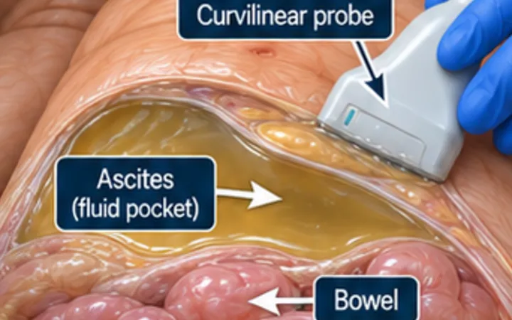
Ultrasound finds even small volumes of ascites, and it shows what the fluid looks like. Clear, free fluid fits the common causes: cirrhosis, heart failure and low albumin. Fluid with echoes, strands or separate pockets has a different list: infection from a perforation, blood, cancer and tuberculosis. The appearance guides the next test. It does not replace it.
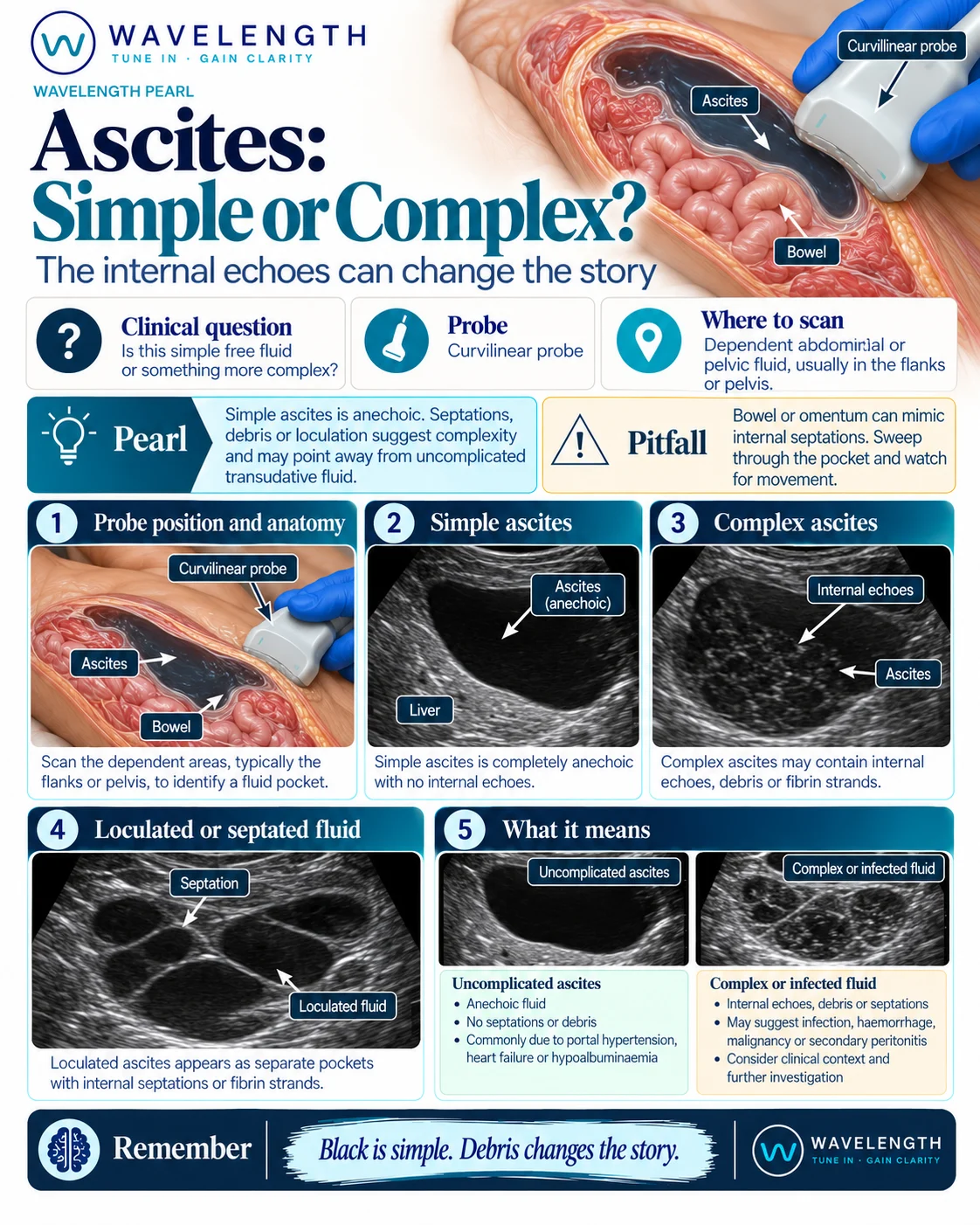
Scan images in the graphic are illustrations, not patient scans.
The scan
- Use the curvilinear probe with the patient supine.
- Look in the dependent places first: around the liver and in Morison's pouch, around the spleen, both flanks and the pelvis.
- In each pocket, turn up the gain a little and look for echoes, strands and walls.
- Watch for movement: free fluid shifts with position and bowel floats in it.
- Look at the liver surface, the spleen and the bowel wall for a cause.
Reading the view
- Simple ascites: black fluid with no echoes, free between bowel loops, which float centrally.
- Complex ascites: low-level echoes, debris or fine strands. Think of infection, blood or malignancy.
- Loculated ascites: separate pockets divided by septations, which do not move freely. Think of infection, tuberculosis, malignancy or previous surgery.
- Clues to a cause: a small, nodular liver suggests cirrhosis; a thick or matted omentum suggests malignancy or tuberculosis.
Action
New ascites, and ascites in anyone admitted to hospital, needs a diagnostic tap, whatever it looks like. Complex or loculated fluid in an unwell patient needs senior review, cross-sectional imaging and a search for the source. See the paracentesis pearl for choosing a safe pocket.