Wavelength Pearls
Abdominal and pelvic ultrasound.
Free fluid, the gallbladder and bile duct, kidneys, bladder, stomach, bowel and the pelvis, in adults and children.
Point-of-care ultrasound of the abdomen and pelvis answers focused questions at the bedside: is there free fluid, is the bile duct dilated, is the kidney obstructed, is the bladder full, is the bowel obstructed, and is there an intrauterine pregnancy?
These pearls cover eFAST, the gallbladder and common bile duct, hydronephrosis and pyonephrosis, the bladder, the stomach, small bowel obstruction, appendicitis, intussusception in children, and pelvic pain in women, including ectopic pregnancy and ovarian torsion.
17 pearls
One technique at a time.
Renal
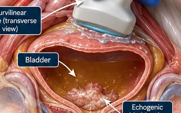
Bladder filling defect: move it, then add colour
Something bright sits in the bladder. Roll the patient and add colour Doppler. Clot moves and has no internal flow. A lesion stuck to the wall stays put and may show vessels.
Abdomen
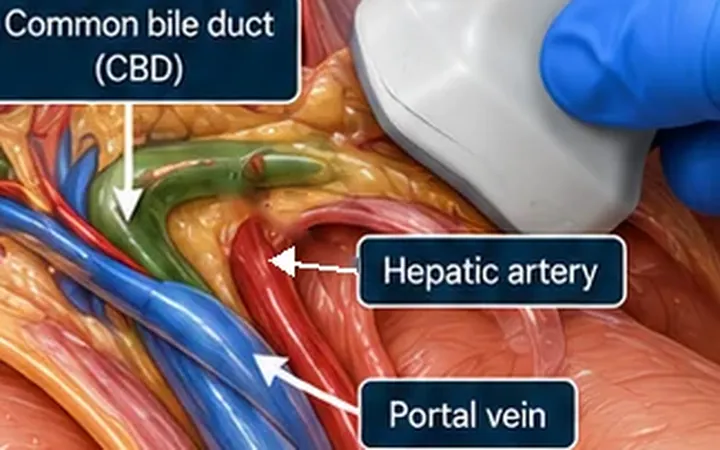
Common bile duct: find the parallel channel
Next to the portal vein lie two tubes, the hepatic artery and the bile duct. Add colour Doppler. The tube with no flow is the duct.
Pelvis
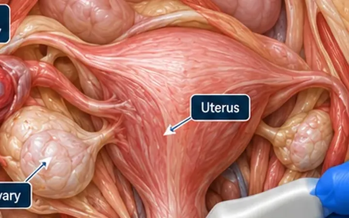
Ectopic pregnancy: empty uterus? Find the adnexa
A positive pregnancy test and pain. The bedside scan has one job first, to find a pregnancy inside the uterus. If you cannot, look beyond it, at the adnexa, the pouch of Douglas and the right upper quadrant.
Abdomen
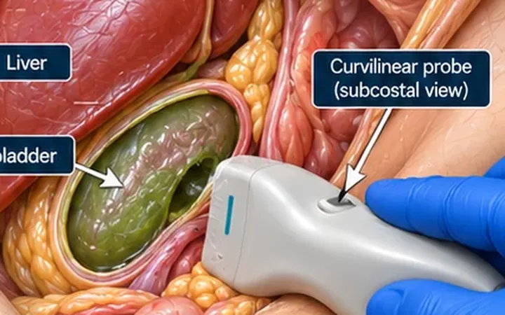
Gallbladder wall: thick does not equal cholecystitis
A gallbladder wall over 3 mm is a clue, not a diagnosis. Look for stones, focal tenderness, distension and pericholecystic fluid before you call cholecystitis.
Pelvis
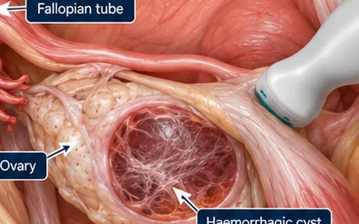
Haemorrhagic cyst: fishnet, not flesh
Lacy internal echoes and a clot with a concave edge and no flow inside it point to a haemorrhagic ovarian cyst. A solid part with blood flow does not, and needs formal imaging.
Pelvis
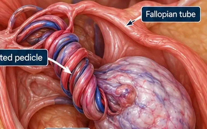
Ovarian torsion: follow the pedicle
Normal Doppler flow does not exclude torsion. Look for an enlarged ovary with peripheral follicles, and follow the vascular pedicle for a whirlpool. Clinical suspicion drives the referral.
Pelvis
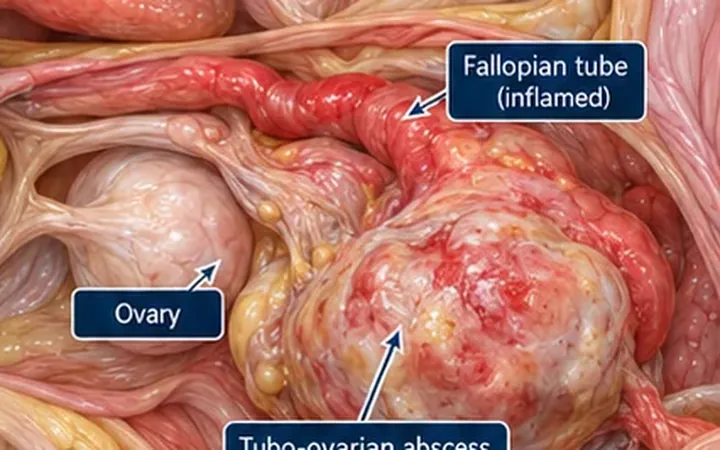
Tubo-ovarian abscess: think complex, thick and tender
Pelvic pain with fever and a complex, thick-walled, tender adnexal mass should raise concern for a tubo-ovarian abscess. POCUS raises the alarm. Formal imaging and gynaecology confirm it.
Renal
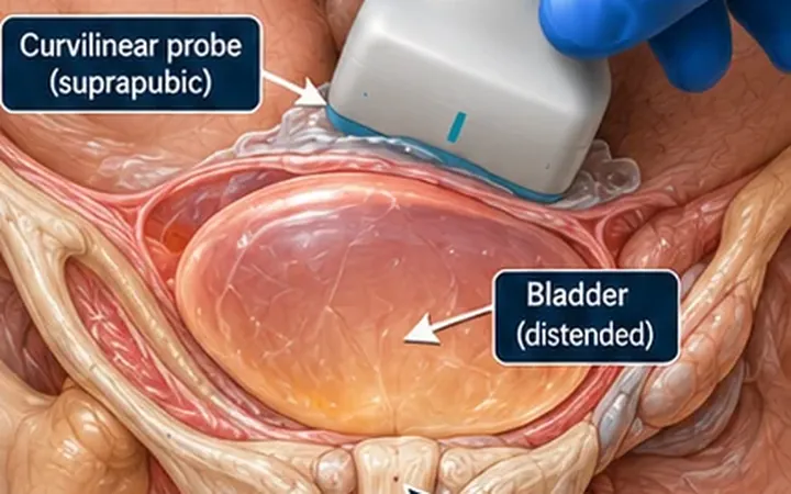
Urinary retention: measure the bladder, then look upstream
Measure the bladder in two planes to confirm it is full. Then scan both kidneys, because a large bladder with hydronephrosis means the obstruction has reached the upper tracts.
Abdomen
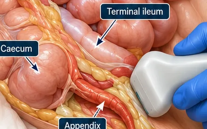
Appendix ultrasound: a positive scan helps
A blind-ending, non-compressible tube over 6 mm across in the right lower quadrant supports appendicitis. Not seeing the appendix does not exclude it. Learn graded compression, what to measure, and how to act on each result.
eFAST
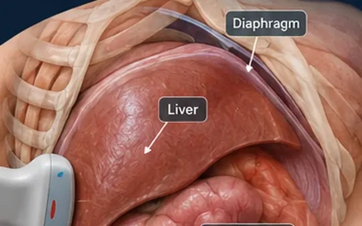
eFAST: look at the liver tip first
In the supine patient, free fluid in the right upper quadrant often collects first at the inferior tip of the liver. Scan the whole view, not just Morison's pouch.
Abdomen
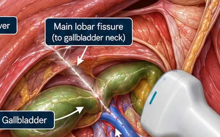
Gallbladder: follow the main lobar fissure
Can't find the gallbladder? Find the portal vein, then follow the bright line running from it. The main lobar fissure leads straight to the gallbladder neck.
Abdomen
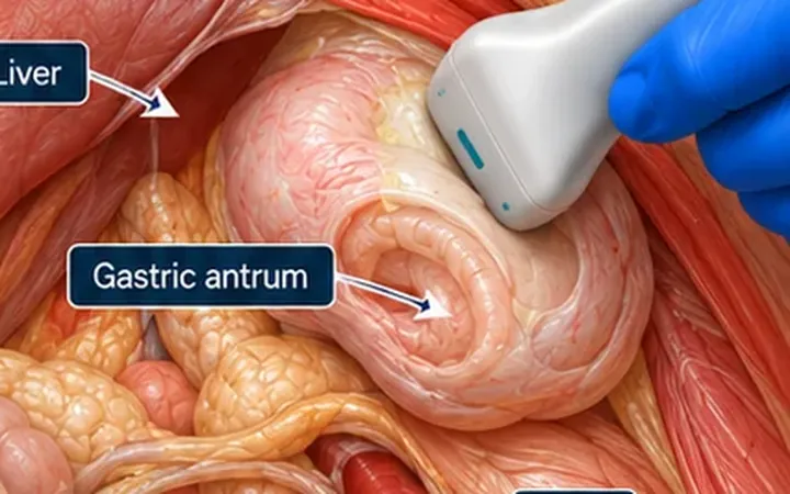
Gastric POCUS: empty, fluid or solid?
Before sedation, one view of the gastric antrum tells you whether the stomach is empty, holds clear fluid, or holds solid food. Answer that question first.
Renal
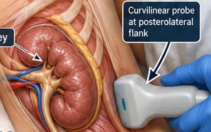
Hydronephrosis: black that branches
True hydronephrosis forms connected, branching black spaces running from the renal pelvis into the calyces. Cysts stay round and separate. Sweep the whole kidney before you decide.
Abdomen
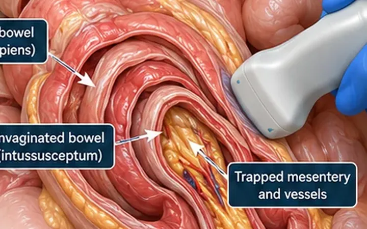
Intussusception: target, then sandwich
A child with colicky pain, drawing up the legs and periods of calm needs a look at the right side of the abdomen. A target in transverse and a sandwich in long axis make the diagnosis. Two planes, one answer.
Renal
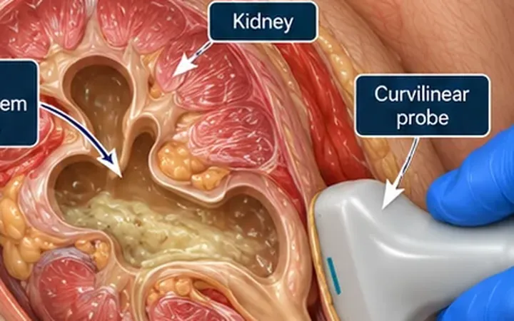
Pyonephrosis: hydronephrosis with dirty echoes
Hydronephrosis in a septic patient is an obstructed, infected kidney until proven otherwise. Debris, layering echoes or a fluid-debris level in the collecting system should make you think of pus and call urology now.
Renal
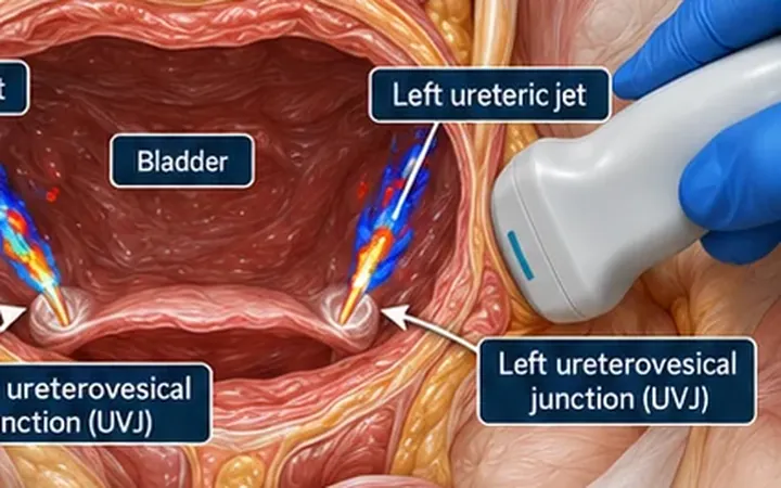
Renal colic: listen with colour
A ureteric jet is urine squirting into the bladder. Colour Doppler shows it. A jet missing on the painful side, with hydronephrosis on that side, supports an obstructing stone. Wait, watch and compare both sides.
Abdomen
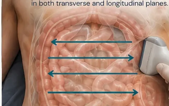
Small bowel obstruction: follow the to-and-fro
Dilated, fluid-filled small bowel with contents moving back and forth without getting anywhere suggests mechanical obstruction. Follow the dilated loops until they collapse to find the transition point.
Practise it with us
One instructor to every four or five delegates, FAMUS-accredited instructors and RCEM-approved ultrasound supervisors, and long, supervised time on the probe.
Newsletter
Get the next one by email.
One email a week: a practical scanning skill, new Learn posts and course dates before they go public.