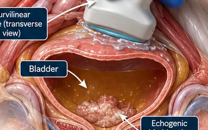
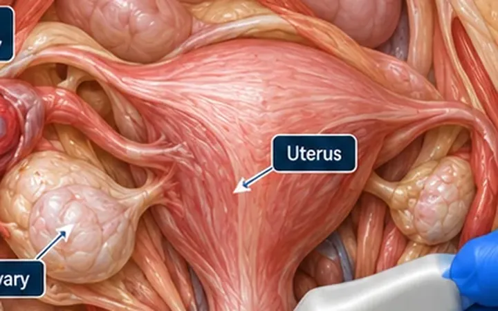
A young woman with sudden pelvic pain and a complex ovarian cyst on scan: is it blood or tissue? Bleeding into a corpus luteum is common and usually settles. Its appearance changes as the blood clots and retracts, and it often mimics a solid mass.
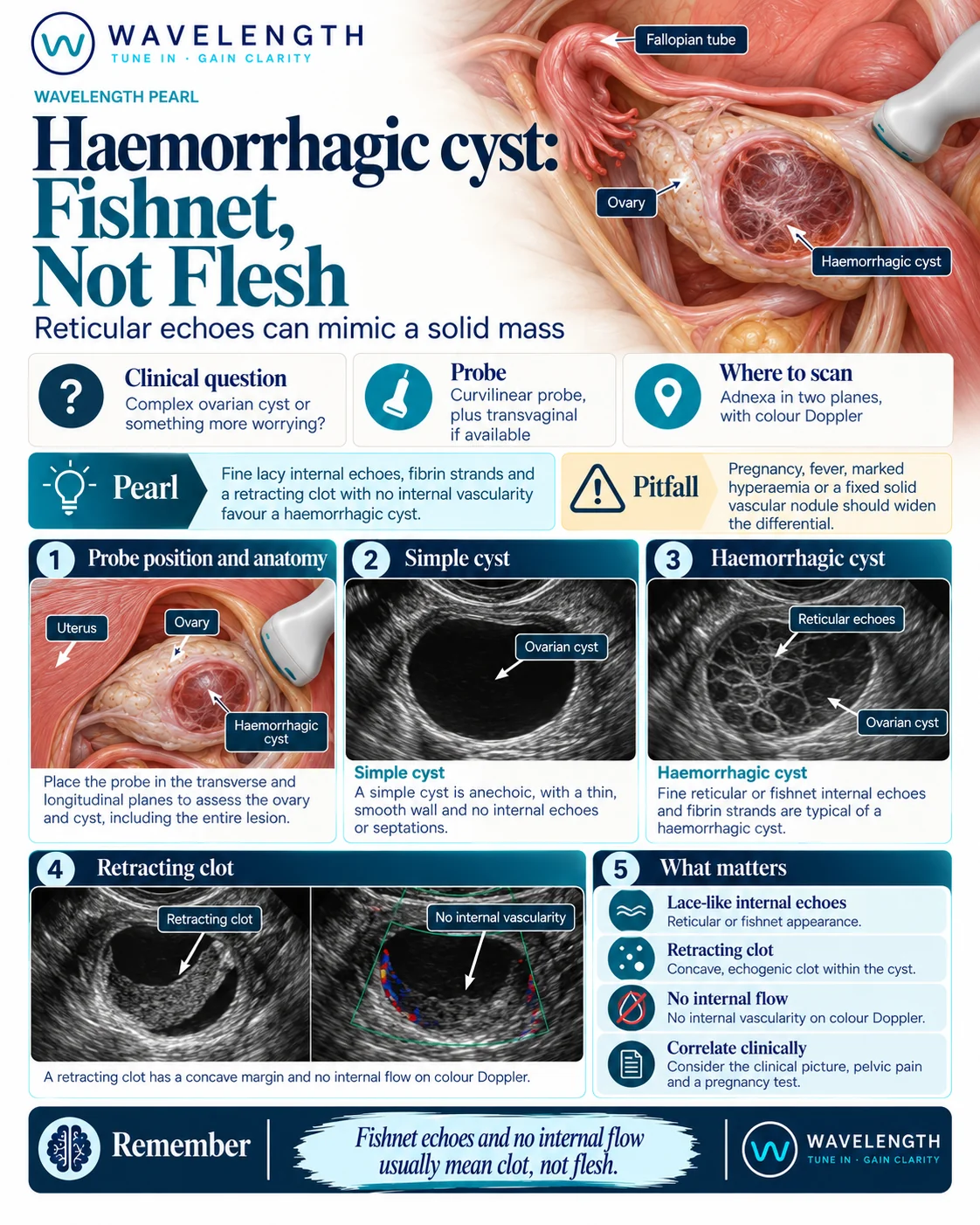
Scan images in the graphic are illustrations, not patient scans.
The scan
- Confirm the pregnancy test result.
- Scan the uterus and both adnexa with the curvilinear probe, in two planes. Transvaginal scanning by a trained operator gives a clearer view.
- Find the cyst and sweep through all of it.
- Look at the internal echoes and any solid-looking part.
- Add colour Doppler with a low velocity scale over any solid-looking area.
- Check the pouch of Douglas and the right upper quadrant for free fluid.
Reading the view
- Simple cyst: black, thin-walled, smooth, with no internal echoes.
- Haemorrhagic cyst: fine, lacy, net-like internal strands with no flow on colour Doppler.
- Retracting clot: a grey mass inside the cyst with a concave or straight edge, no flow inside it, and sometimes moving with the patient.
- Ruptured cyst: echogenic free fluid around the ovary or in the pouch of Douglas.
What to do next
A typical haemorrhagic cyst in a stable, non-pregnant patient usually needs analgesia and follow-up according to local guidance. Large haemoperitoneum, haemodynamic instability or an atypical appearance need urgent gynaecology review.