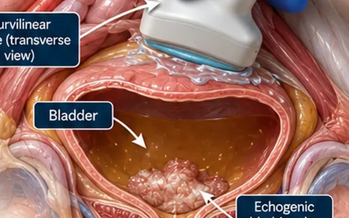
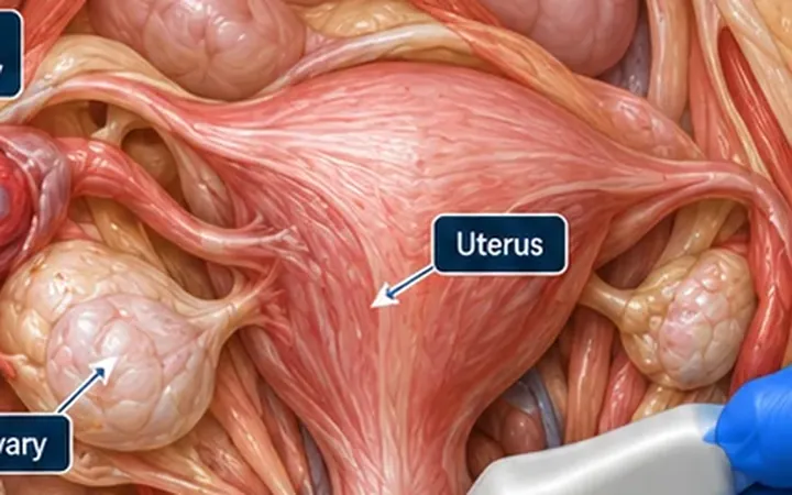
Pelvic inflammatory disease often looks like little on ultrasound. When it forms an abscess, the scan changes, and a complex mass beside the uterus becomes the clue to pelvic sepsis.
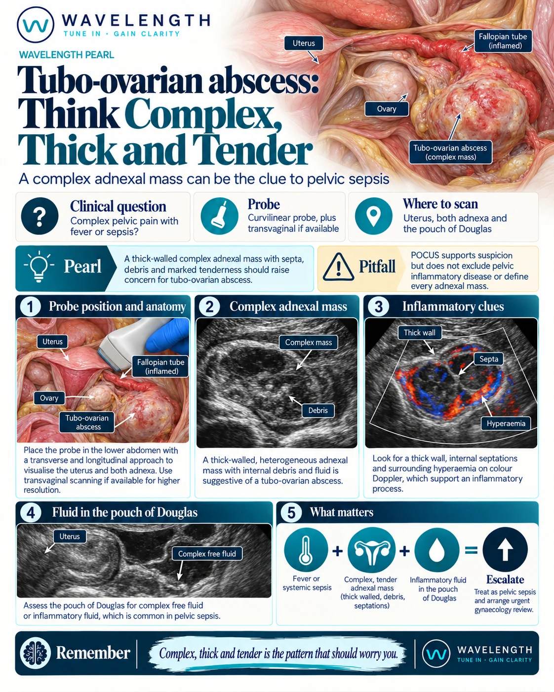
Scan images in the graphic are illustrations, not patient scans.
The scan
- Confirm the pregnancy test result.
- Scan transabdominally with the curvilinear probe, in transverse and sagittal, covering the uterus and both adnexa. Transvaginal scanning by a trained operator gives a clearer view.
- Look beside the uterus for a mass. Note its wall, its contents and whether you can still see a normal ovary.
- Add colour Doppler around the mass.
- Press gently over it with the probe and note tenderness.
- Check the pouch of Douglas for fluid.
Reading the view
- Tubo-ovarian abscess: a thick-walled, multilocular adnexal mass with internal debris, septa or fluid levels, often with the ovary no longer separate from the tube.
- Inflammation: hyperaemia in the wall and septa on colour Doppler.
- Pyosalpinx: a dilated, fluid-filled tube with thick walls and debris.
- Free fluid: complex fluid in the pouch of Douglas.
What to do next
Treat as pelvic sepsis: blood cultures, intravenous antibiotics according to local guidance, and urgent gynaecology review. Large abscesses or those not responding to antibiotics need drainage. Arrange formal imaging to confirm the diagnosis.