Fasting times guide sedation and anaesthesia, but they do not tell you what is in the stomach. Trauma, pain, opioids, diabetes and pregnancy all slow gastric emptying. A quick scan of the gastric antrum shows the contents directly.
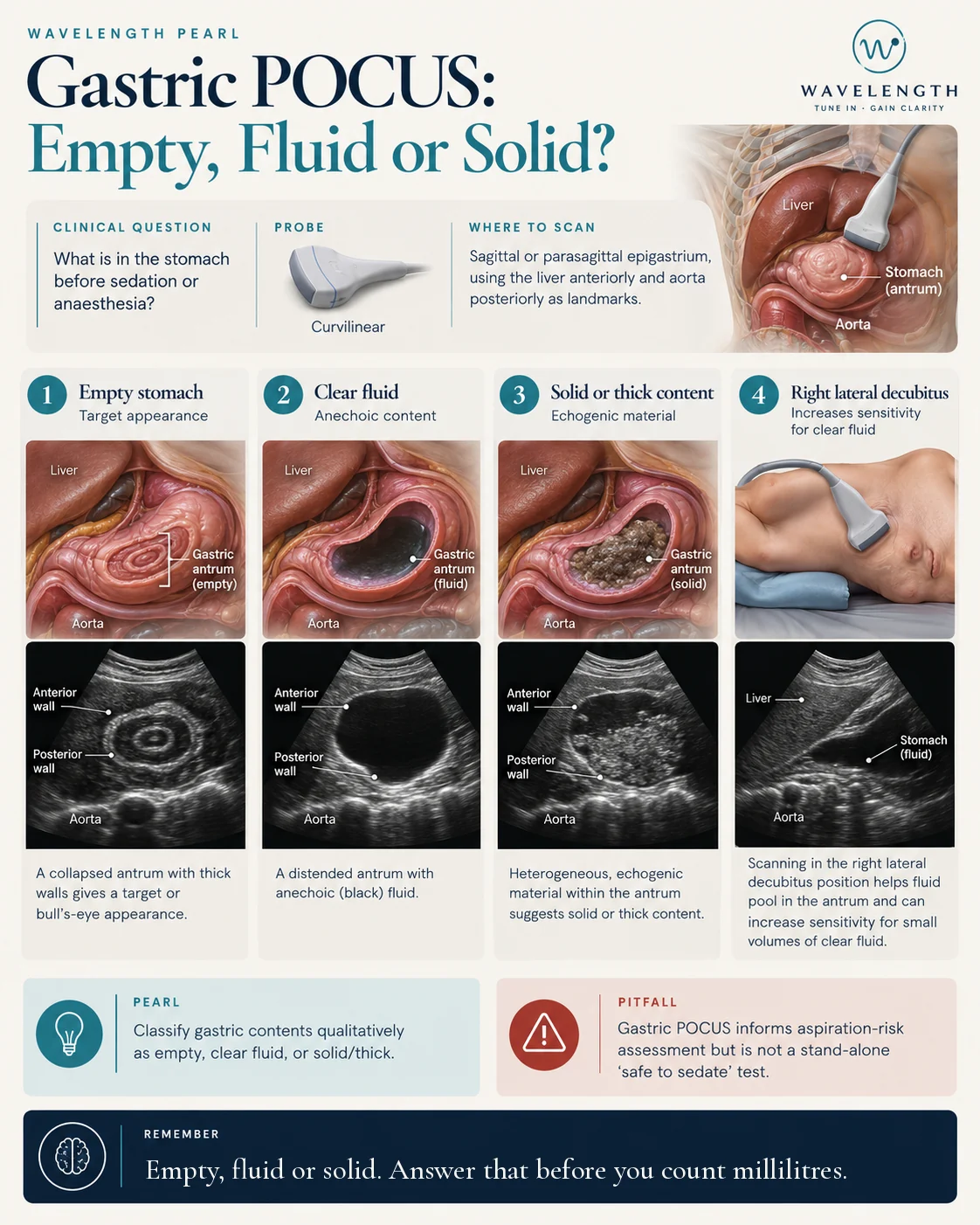
Scan images in the graphic are illustrations, not patient scans.
The scan
- Use the curvilinear probe.
- Place it sagittally in the epigastrium, just to the right of the midline, marker to the head.
- Find the landmarks: the left lobe of the liver at the top of the screen, and the aorta or IVC deep to the stomach.
- The gastric antrum lies between them.
- Scan first with the patient supine, then in the right lateral decubitus position, right side down. Fluid runs into the antrum in this position, so it shows small volumes.
Reading the view
- Empty. A small, collapsed antrum with layered walls, like a target or bull's-eye.
- Clear fluid. A distended, round antrum with black, anechoic contents.
- Solid or thick. Mixed, bright contents. Fresh solid food often looks like frosted glass, which blurs the back wall.
Taking it further
When the antrum holds clear fluid, the cross-sectional area measured in the right lateral position estimates the volume. Validated formulas exist for adults. Learn qualitative assessment first, because it answers the main question.