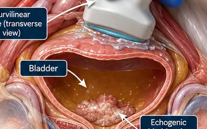
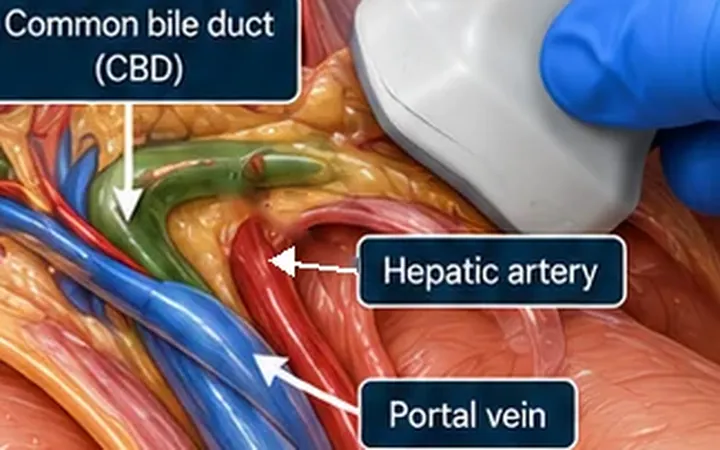
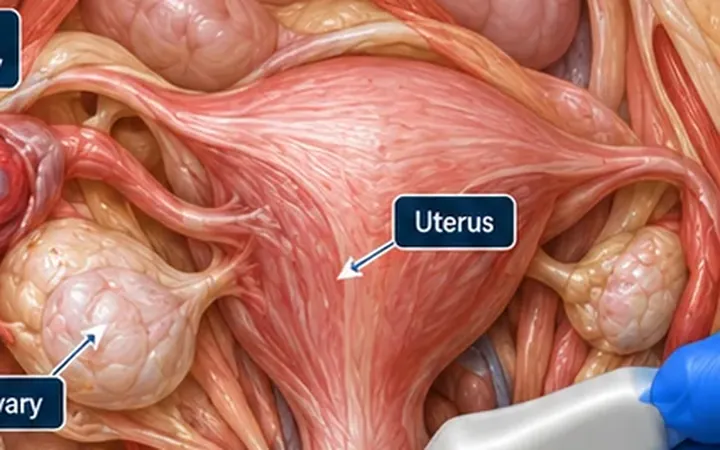
A thick gallbladder wall catches the eye and tempts a quick diagnosis of cholecystitis. Heart failure, ascites, low albumin, liver disease and a recent meal all thicken the wall without any inflammation of the gallbladder. The wall tells you something is going on. The pattern around it tells you what.
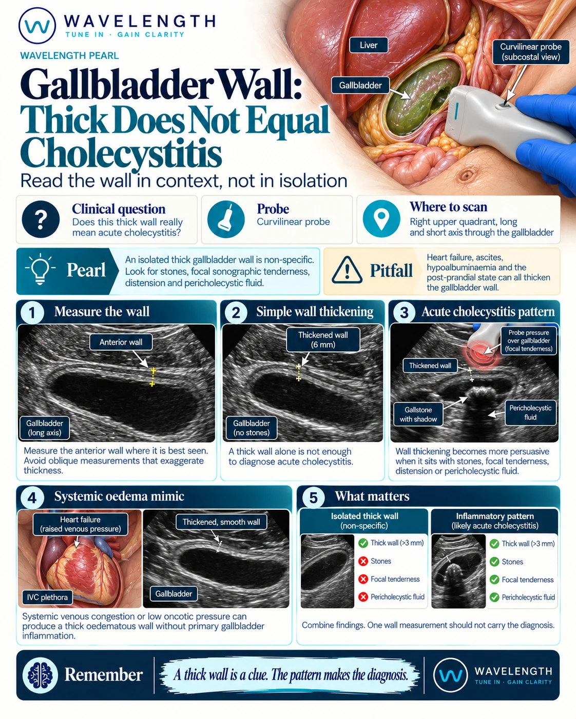
Scan images in the graphic are illustrations, not patient scans.
The scan
- Find the gallbladder in long axis with the curvilinear probe, below the right costal margin or between the lower ribs.
- Measure the anterior wall, the one nearest the probe, with the beam at 90° to the wall. The posterior wall blends with bowel behind it, and an oblique cut makes any wall look thicker.
- Sweep the whole gallbladder in long and short axis, from fundus to neck, looking for stones.
- Press over the gallbladder with the probe and ask where it hurts most.
- Look around the gallbladder for a rim of fluid, and check its size.
Reading the view
- Thickened wall: over 3 mm.
- Supports acute cholecystitis: gallstones, especially one stuck in the neck, a positive sonographic Murphy's sign (maximal tenderness under the probe over the gallbladder), a distended gallbladder and pericholecystic fluid.
- Points elsewhere: a smooth, layered thick wall with no stones and no focal tenderness, in a patient with heart failure, ascites, low albumin, liver disease or a recent meal.
What the scan does not tell you
Acalculous cholecystitis occurs in critically ill patients without stones, and a gangrenous gallbladder loses its tenderness. When the picture does not fit, use bloods, the clinical course and formal imaging.