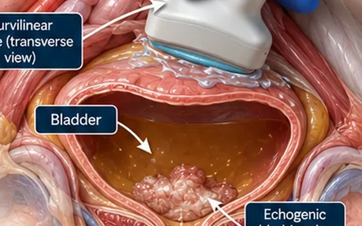
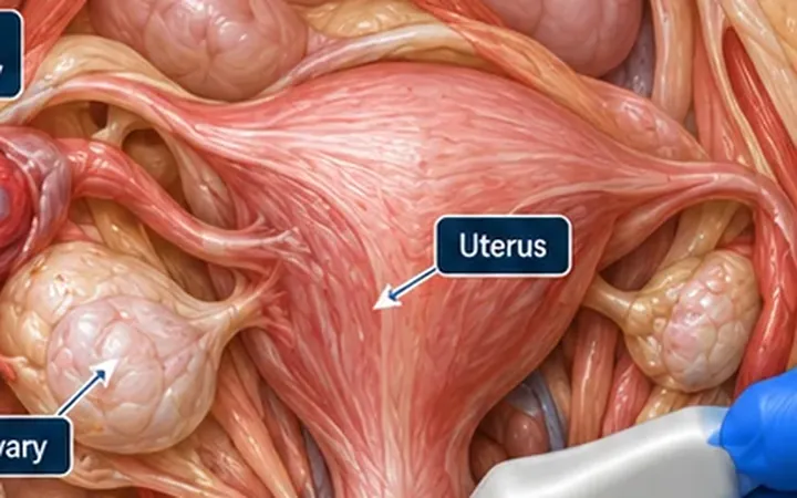
Ovarian torsion threatens the ovary within hours. Many clinicians look for absent Doppler flow, but the ovary has two blood supplies and torsion is often partial or intermittent, so flow is frequently present. The twisted pedicle and the shape of the ovary tell you more.

Scan images in the graphic are illustrations, not patient scans.
The scan
- Confirm the pregnancy test result.
- Scan transabdominally with the curvilinear probe through a full bladder if possible. Transvaginal scanning by a trained operator gives a clearer view.
- Find the uterus, then each ovary beside it. Find both ovaries before you call one abnormal.
- Compare their size and appearance.
- Look for the pedicle, the vessels running from the ovary towards the uterus and pelvic side wall. Follow it in short axis and watch for a swirl.
- Add colour Doppler, but read it last.
Reading the view
- Enlarged ovary: clearly larger than the other side, often with oedematous central tissue and follicles pushed to the edge.
- Whirlpool sign: the pedicle twists into a spiral or target. This is the most specific sign.
- Associated mass: a cyst or dermoid often acts as the lead point.
- Free fluid: a small amount around the ovary is common.
- Doppler: reduced or absent venous flow supports torsion. Arterial flow is often still present.
What to do next
Sudden, severe, one-sided pelvic pain with nausea or vomiting needs urgent gynaecology review, whatever the scan shows. The diagnosis is confirmed at surgery, and delay risks the ovary. In children and adolescents, involve the paediatric surgical or gynaecology team early.