A diagnostic tap is part of the assessment of anyone admitted with ascites, and a large-volume drain relieves pain and breathlessness. Most complications come from the needle path: bowel caught against the wall, or a bleed from an abdominal wall vessel. Ultrasound shows both before you start. In large observational studies, ultrasound guidance was associated with fewer bleeding complications.
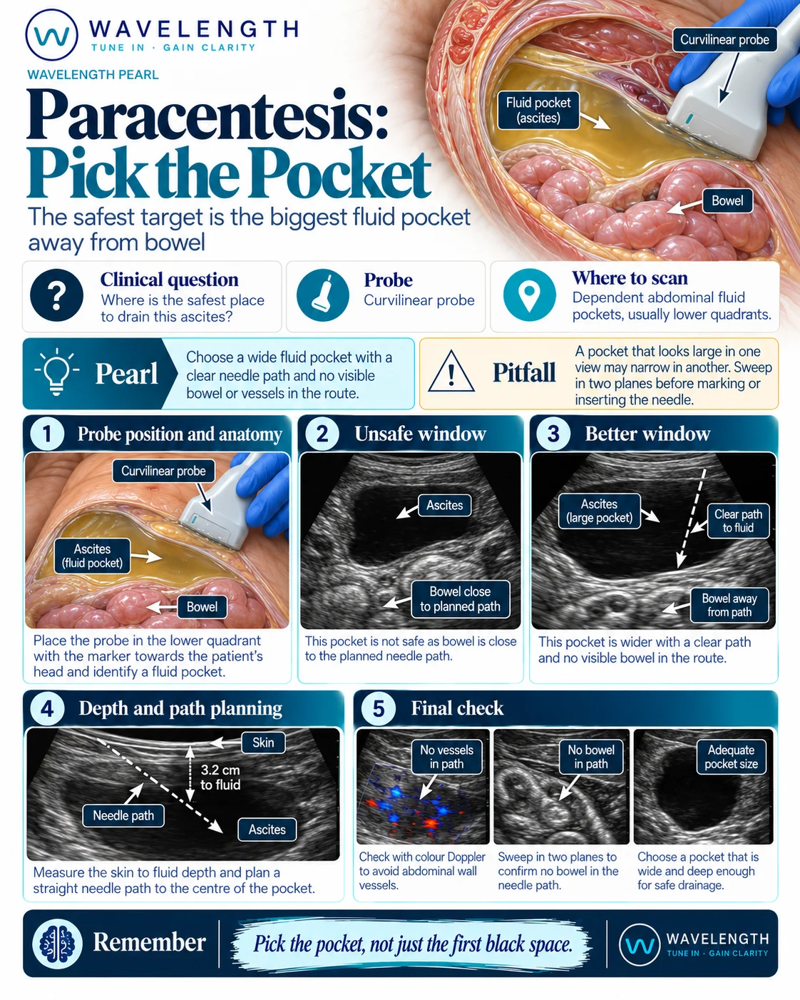
Scan images in the graphic are illustrations, not patient scans.
The scan
- Lay the patient supine, tilted slightly towards the side you plan to drain.
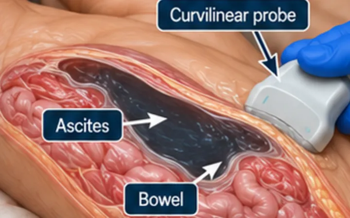
- Use the curvilinear probe in both lower quadrants, lateral to the rectus muscle. The left lower quadrant is often deeper and has thinner fat.
- Find the largest pocket and sweep through it in two planes. Look for bowel loops floating up into the path.
- Switch to a linear probe and colour Doppler over the planned entry point to find the inferior epigastric vessels and any wall varices.
- Measure the depth from skin to fluid, and the depth of fluid under that point.
- Mark the site and drain without moving the patient, or drain under real-time guidance.
Reading the view
- Good window: a deep pocket, several centimetres of fluid, with bowel well away from the path through the breathing cycle.
- Unsafe window: bowel loops close to the wall, a shallow pocket, or a vessel on colour Doppler in the path.
- Fluid character: clear black fluid, or debris and septations, which change the differential. See the ascites pearl.
Action
Follow your local paracentesis procedure and consent process. Routine correction of clotting is not usually needed before paracentesis in cirrhosis, but follow local guidance. Send fluid for cell count, culture and albumin; a neutrophil count over 250 per cubic millimetre suggests spontaneous bacterial peritonitis.