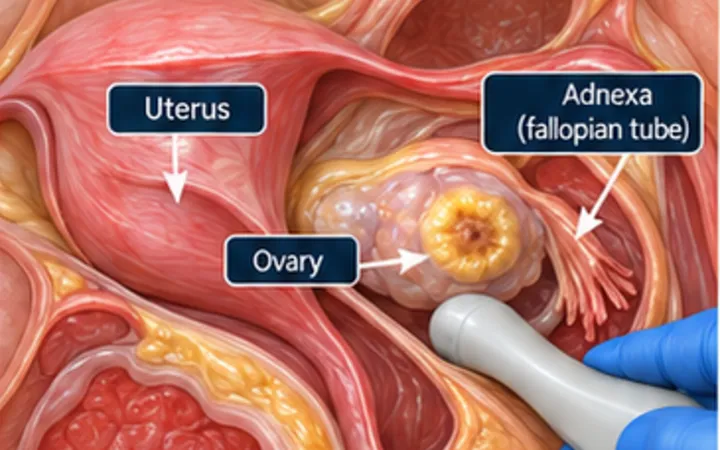
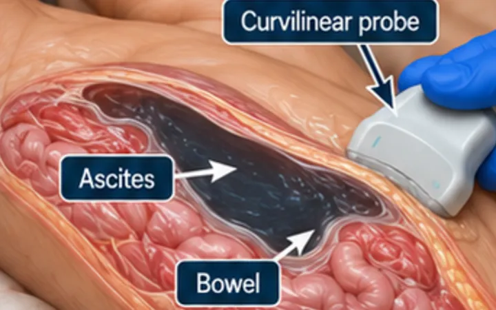
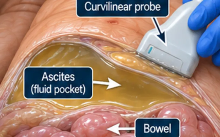
Left iliac fossa pain with fever and tenderness in an older adult is often diverticulitis. CT is the usual test when complications are suspected, but in experienced hands ultrasound has shown accuracy close to CT, and it can be done at once, without radiation. The key is to put the probe exactly on the point of tenderness.
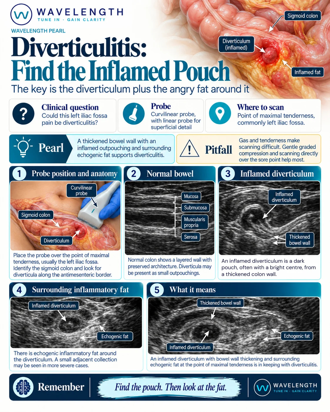
Scan images in the graphic are illustrations, not patient scans.
The scan
- Ask the patient to point with one finger to where it hurts most.
- Start with the curvilinear probe, then change to a linear probe for detail.
- Use graded compression: press slowly and steadily to push bowel gas out of the way.
- Find the colon, with its layered wall and gas, and follow it through the tender area in long and short axis.
- Look at the wall, the pouches arising from it and the fat around it. Then look for fluid, a collection or gas outside the bowel.
Reading the view
- Normal colon: a thin layered wall, often with small, quiet diverticula.
- Thickened wall: a segment of colon wall thicker than about 4 to 5 mm at the tender point.
- Inflamed diverticulum: a small dark pouch arising from the wall, often with a bright centre from gas or a faecolith.
- Inflamed fat: bright, non-compressible fat around the pouch, the most visible sign.
- Complications: a collection with debris suggests an abscess; bright foci with dirty shadowing outside the bowel suggest free gas.
Action
Follow your local pathway. Patients who are unwell, immunosuppressed, or have an abscess, free gas or uncontrolled pain need senior surgical review and CT. Make sure a cancer is excluded after the episode, as your local pathway advises, especially if the diagnosis was made on imaging alone.