In early pregnancy, almost every woman has a corpus luteum: a thick-walled cyst inside the ovary, often with a ring of blood flow around it. A tubal ectopic pregnancy can also look like a ring. The key is position. This pearl follows on from the ectopic pregnancy pearl, which starts with the uterus.
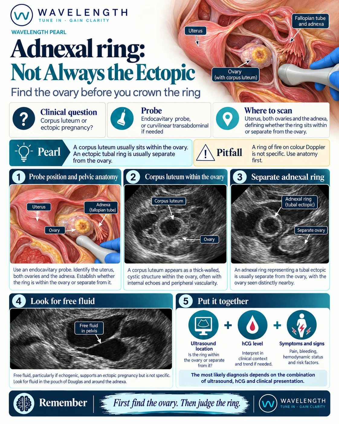
Scan images in the graphic are illustrations, not patient scans.
The scan
- Explain the scan, offer a chaperone and record the offer. Confirm the pregnancy test first.
- Look for an intrauterine pregnancy, as in the ectopic pearl.
- Find each ovary beside the uterus: an oval structure with small follicles.
- Find the ring and decide whether it is inside the ovary or separate from it.
- With gentle pressure from the probe, watch whether the ring moves with the ovary or separately from it.
- Look for free fluid in the pouch of Douglas and around the liver.
Reading the view
- Corpus luteum: inside the ovary, surrounded by ovarian tissue, with a thick wall that is usually less bright than the endometrium. It moves with the ovary.
- Tubal ectopic: a ring separate from the ovary, often brighter than the ovary, sometimes containing a yolk sac or fetal pole. It moves separately from the ovary.
- Free fluid: echogenic fluid in the pelvis or fluid around the liver raises concern for rupture.
Action
An unstable patient with a positive test and free fluid needs resuscitation and immediate gynaecology review. Otherwise, a positive test with no definite intrauterine pregnancy goes to the early pregnancy unit for a transvaginal scan and serial hCG, with clear safety-netting.