Acute mesenteric ischaemia is uncommon, lethal and easily missed. An embolus to the superior mesenteric artery, usually from atrial fibrillation, is the classic cause; thrombosis on an atheromatous artery, low-flow states and mesenteric venous thrombosis also cause it. The early abdomen can be soft while the pain is severe. Mortality climbs the longer it goes unrecognised, so the diagnosis has to be considered before the bloods or the scan turn abnormal.
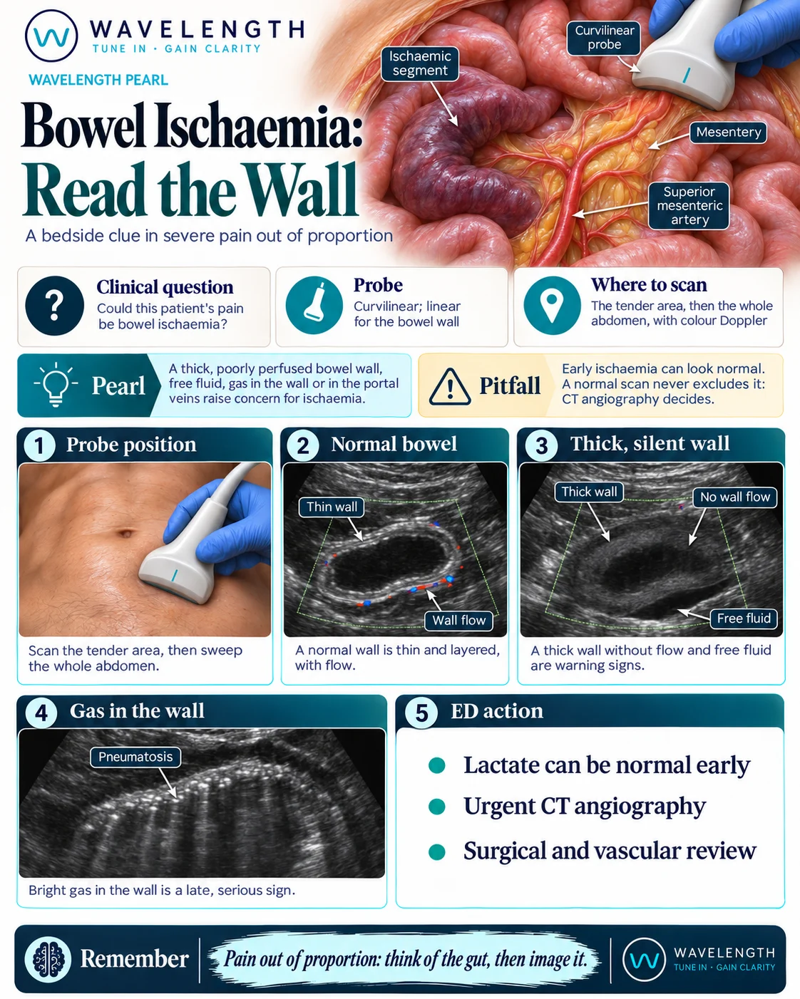
Scan images in the graphic are illustrations, not patient scans.
The scan
- Use the curvilinear probe to sweep the whole abdomen, starting at the tender area, and look for free fluid.
- Switch to the linear probe to see the layers of the bowel wall in a thinner patient.
- Measure the wall of any abnormal loop.
- Add colour Doppler with a low velocity scale, and compare the wall flow with a normal loop.
- Look in the liver for gas in the portal veins.
Reading the view
- Normal bowel: a thin, layered wall, usually under 3 mm, with some flow in it and visible peristalsis.
- Worrying loop: a thick wall, little or no flow, reduced peristalsis and free fluid beside it.
- Late signs: bright gas in the bowel wall with dirty shadowing (pneumatosis), or gas in the portal veins.
- Also consider: obstruction with strangulation, which can look similar.
Action
Resuscitate, give analgesia and broad-spectrum antibiotics, and call the surgeons and vascular team early. Request urgent CT angiography, and do not wait for the creatinine if the picture fits. Anticoagulation and the route to revascularisation are decided with the specialist teams.