A catheter that has stopped draining is a common call in the emergency department and on the wards. Flushing it blind treats only one of the possible causes, and it does nothing for a balloon in the urethra or a patient who is making no urine. Ultrasound shows where the balloon is and how much urine lies around it.
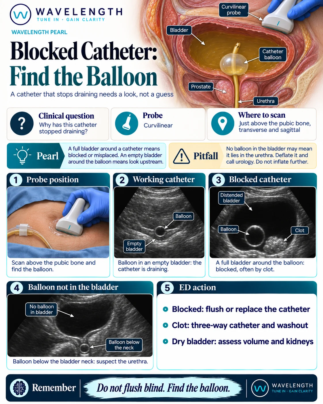
Scan images in the graphic are illustrations, not patient scans.
The scan
- Check the outside first: a kinked tube, a closed valve, or a bag above the bladder.
- Use the curvilinear probe just above the pubic bone, in transverse and sagittal.
- Find the bladder and judge how full it is. Measure the volume if it is large.
- Find the balloon: a round, black circle with a bright wall.
- Look for bright, irregular material beside it. If you find a full bladder or no urine, go on to look at both kidneys.
Reading the view
- Balloon in an empty bladder: the catheter is in place and draining. Look upstream at the patient's volume state and the kidneys.
- Full bladder around the balloon: the catheter is blocked, often by clot or debris.
- Bright material that moves and has no flow: clot in the bladder.
- Full bladder with no balloon inside it: look below the bladder neck. A balloon there lies in the urethra.
Action
If the catheter is blocked, flush it or replace it, following local policy. Clot retention needs a three-way catheter and bladder washout, with urology involved if the bleeding continues. If the bladder is dry, assess the patient's volume state, the kidneys and the blood results rather than changing the catheter. A balloon in the urethra needs deflating and urgent urology review.