The vesicoureteric junction is one of the narrowest points in the ureter, so stones often lodge there. Most of the ureter is hidden by bowel gas, but its lowest part lies just behind the bladder. Fill the bladder and you have a window onto it, which is easy to forget when the scan has stopped at the kidney.
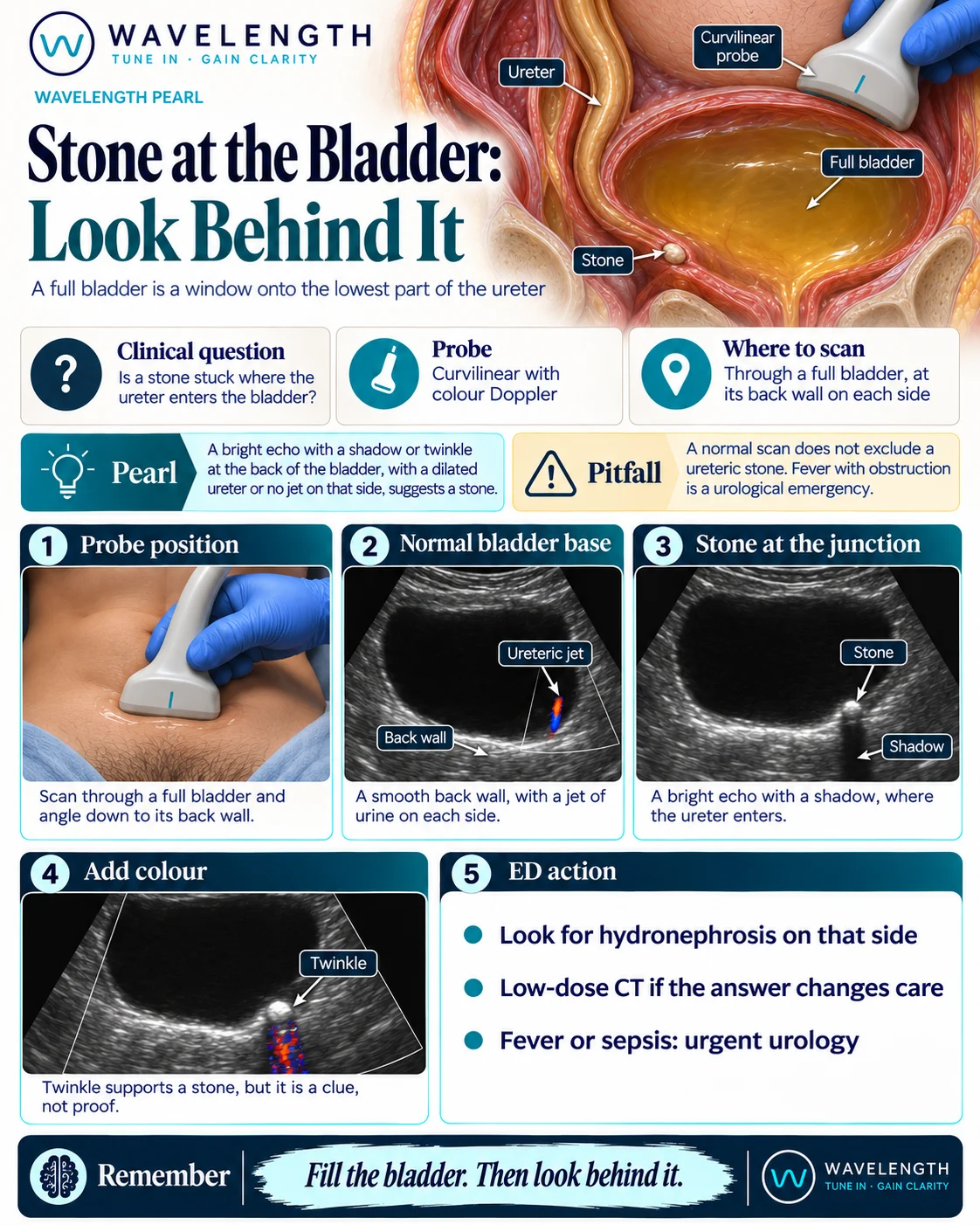
Scan images in the graphic are illustrations, not patient scans.
The scan
- Start with a reasonably full bladder. Ask the patient to drink and come back if it is empty.
- Use the curvilinear probe above the pubic bone, angled down to the back wall.
- Look at the back wall on each side, in transverse and then sagittal.
- Follow any dilated ureter down to the bladder.
- Add colour Doppler, watch for ureteric jets, and look for a twinkle behind any bright echo.
- Scan both kidneys for hydronephrosis.
Reading the view
- Normal: a smooth back wall, and a jet of urine from each side.
- Stone at the junction: a bright echo with a shadow where the ureter enters.
- Supporting signs: a dilated lower ureter, a twinkle, or no jet on the painful side after a few minutes of watching.
- Stone loose in the bladder: it rolls when the patient turns. A stone in the junction stays where it is.
- Bladder mass or clot: a filling defect does not shadow in the same way, and a tumour may have flow in it.
Action
Look for hydronephrosis on that side. Low-dose CT remains the usual test for a suspected stone in adults, where the answer changes care, and ultrasound comes first in pregnancy and in children. Fever or sepsis with a suspected stone needs urgent urology, as do a single kidney, a kidney injury or uncontrolled pain. An older patient with a first episode of renal colic needs the aorta checked too.