A liver abscess presents with fever and right upper quadrant pain, often with few other clues, and is easily mistaken for gallbladder disease. Most are pyogenic, spreading from the biliary tree or from an abdominal source such as appendicitis or diverticulitis. An amoebic abscess can appear months or years after travel. A scan aimed only at the gallbladder can miss it, so sweep the whole liver.
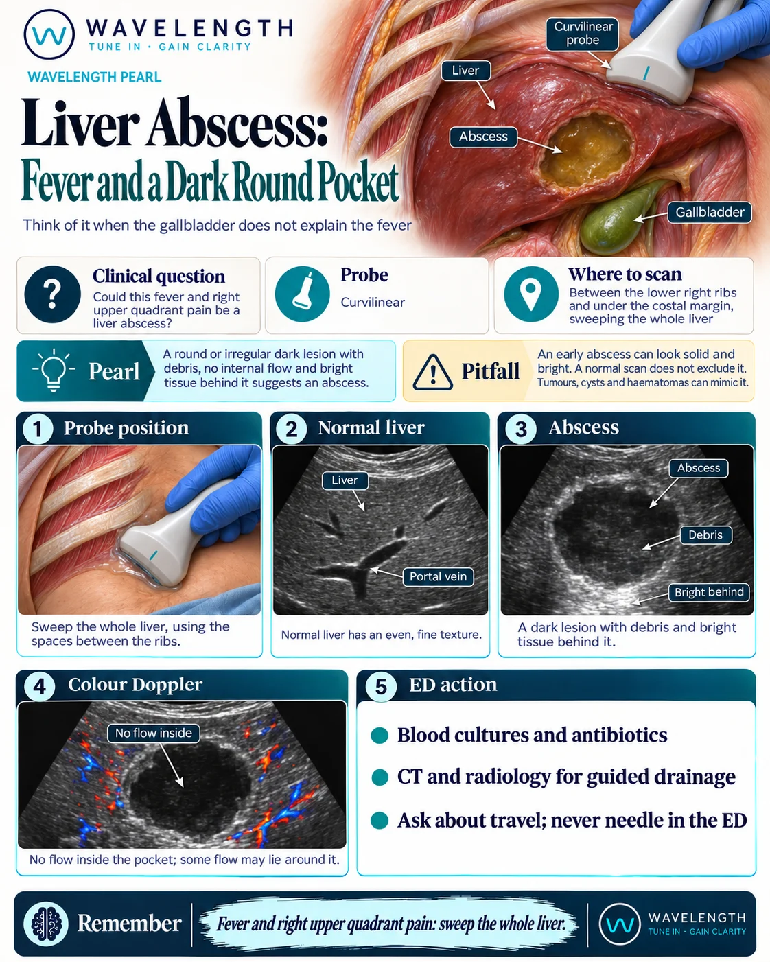
Scan images in the graphic are illustrations, not patient scans.
The scan
- Use the curvilinear probe between the lower right ribs and under the costal margin.
- Sweep the whole liver through each window, including the dome and the left lobe.
- Look at any lesion in two planes, and note its wall, its contents and the tissue behind it.
- Add colour Doppler.
- Look at the gallbladder and the bile ducts for a source, and above the diaphragm for a right pleural effusion.
Reading the view
- Normal liver: an even, fine texture with vessels running through it.
- Abscess: a round or irregular dark lesion with debris and a thick, irregular wall, and brighter tissue behind it.
- Colour Doppler: no flow inside the pocket, though some flow may lie around it.
- Gas in the abscess: bright echoes with dirty shadowing, which is a sign of a more severe infection.
Action
Take blood cultures and start antibiotics according to local guidance. Request CT and speak to radiology about guided drainage. Ask about travel, because amoebic abscesses are treated differently and hydatid disease changes the plan: send serology and take advice. Never needle a liver lesion in the emergency department.