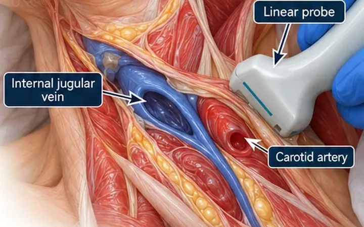
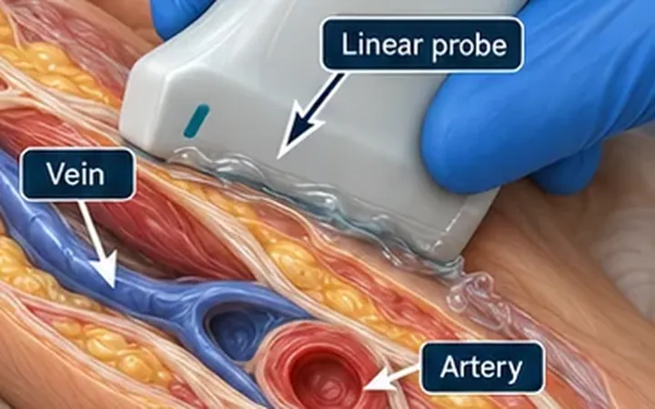
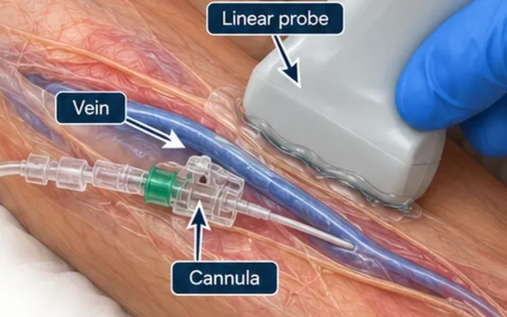
An arterial line in a shocked patient is hard to place by palpation, because the pulse is weak and the artery is small. Ultrasound shows the artery, its walls and its neighbours, so you can choose the easiest stretch instead of the first pulse you feel. A 2014 meta-analysis of randomised trials found that ultrasound guidance improves the chance of success at the first attempt and reduces the number of attempts for radial artery catheterisation.
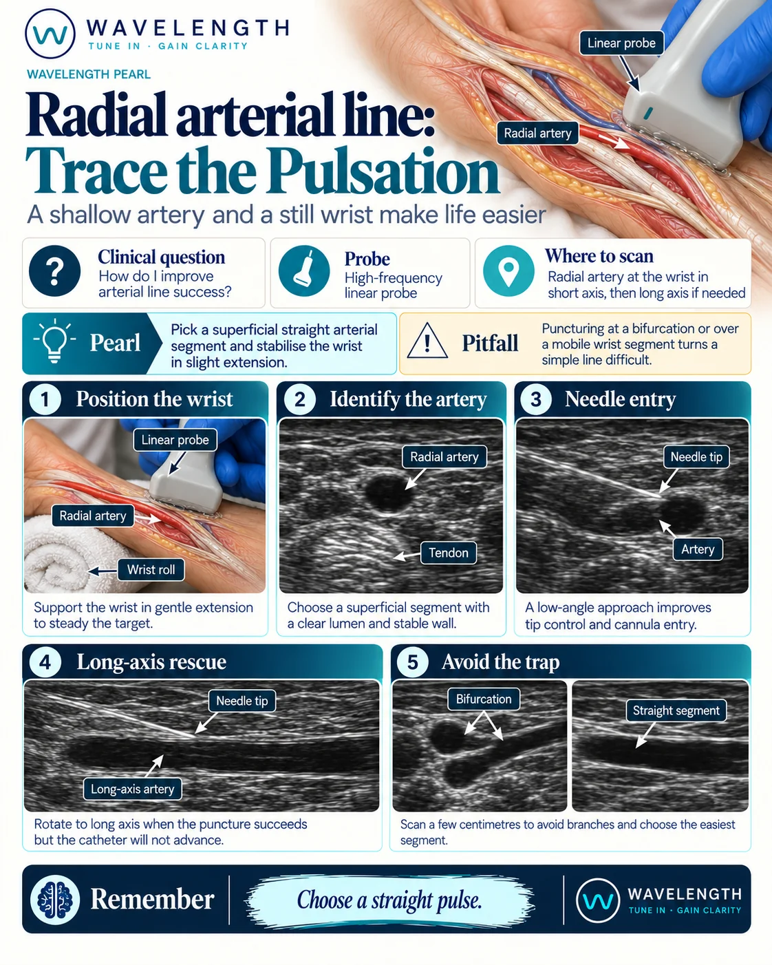
Scan images in the graphic are illustrations, not patient scans.
Before you start
Record consent where possible, check the hand is well perfused and choose the limb according to your trust's policy. Use sterile technique and local anaesthetic in the awake patient.
The scan
- Support the wrist on a roll or folded towel, in gentle extension. Over-extension can flatten the artery, so stop short of the end of range. Keep the hand still.
- Scan the distal forearm in short axis with a high-frequency linear probe. The radial artery is a round, pulsating vessel, close to a tendon and usually with a vein on each side.
- Slide up and down the forearm a few centimetres. Choose a straight, superficial segment with a clear lumen and stable wall, away from branches and bends.
- Enter at a low angle. A shallow angle gives better control of the tip and an easier catheter entry.
- When you see pulsatile blood return, lower the angle and advance, then thread the catheter under vision.
- If the puncture succeeds but the catheter will not advance, rotate to long axis and look at the tip and the wall, as for a peripheral line.
Reading the view
- Radial artery. A round, pulsating, non-compressible lumen with a bright wall.
- Tendon. A bright, fibrillar structure beside the artery that does not pulsate.
- Bifurcation or branch. Two lumens in one segment. Move to a straight stretch.
After the attempt
After you remove a catheter or a failed needle, apply firm pressure over the site until bleeding stops, and check the hand's colour and warmth. Watch for haematoma and for any change in perfusion.