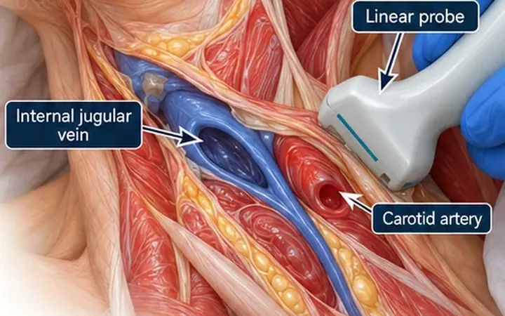
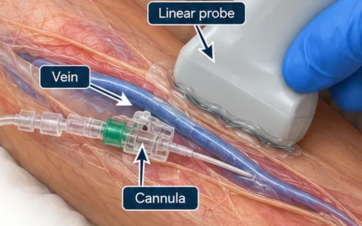
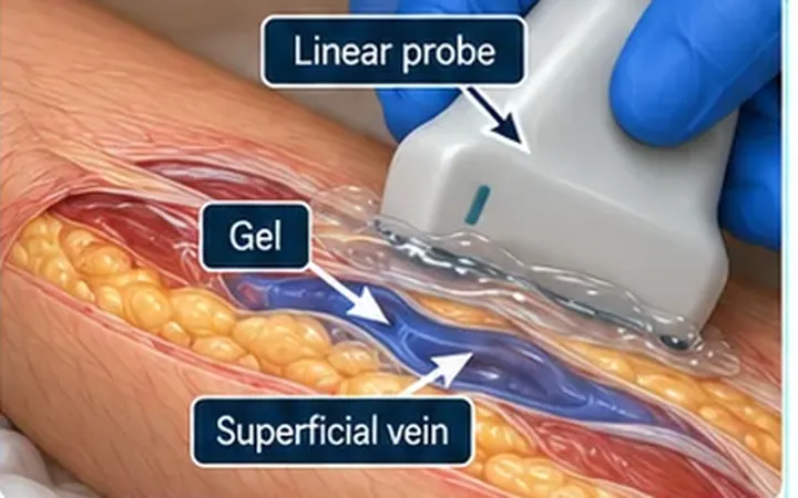
Most failed ultrasound-guided cannulations start before the needle touches the skin. The vein sits at the edge of the screen, or it is flattened by the probe, or the scanner has not checked that the target stays in view as the probe moves. Fixing the picture first costs a few seconds and saves attempts.
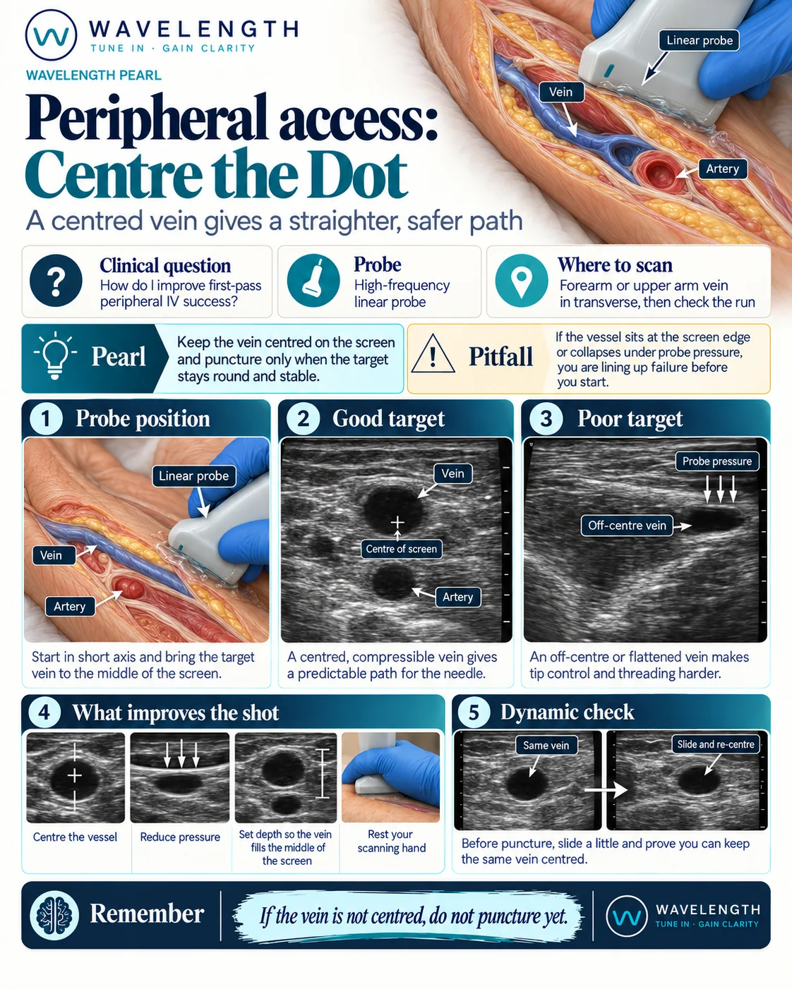
Scan images in the graphic are illustrations, not patient scans.
The scan
- Apply a tourniquet and scan the limb in short axis with a high-frequency linear probe. Find the vein and the artery beside it.
- Slide the probe until the target vein sits in the middle of the screen, under the centre of the probe.
- Reduce the pressure until the vein is round. A flattened vein is smaller than it really is.
- Set the depth so the vein fills the middle of the screen, with the skin and the deeper structures still in view.
- Rest the heel of your scanning hand on the patient, so small movements do not shift the image.
- Run the dynamic check: slide a short way along the vein, then back, and keep the same vein centred and round. If you cannot do this, you will not be able to follow the needle either.
- Puncture only when the target stays round and stable.
Reading the view
- Good target. A round, black, compressible vein in the middle of the screen, with the artery clearly separate.
- Poor target. A vein at the screen edge, or a flat slit under heavy pressure. The needle path is harder to predict and threading is harder.
When ultrasound helps most
A 2022 Cochrane review found that ultrasound guidance probably improves first-pass success in adults with moderately difficult access and may improve it in those with difficult access. In patients with easy access, first-pass success was lower with ultrasound, and the procedure took longer. Use ultrasound when the vein is hard to see or feel, not as a replacement for landmark cannulation in every patient.