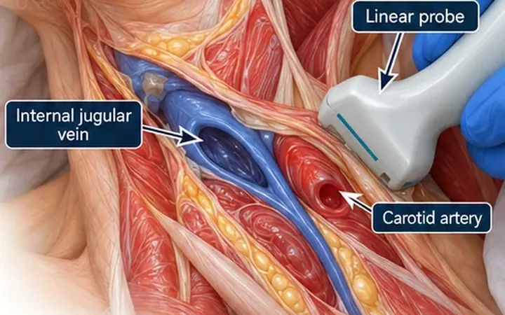
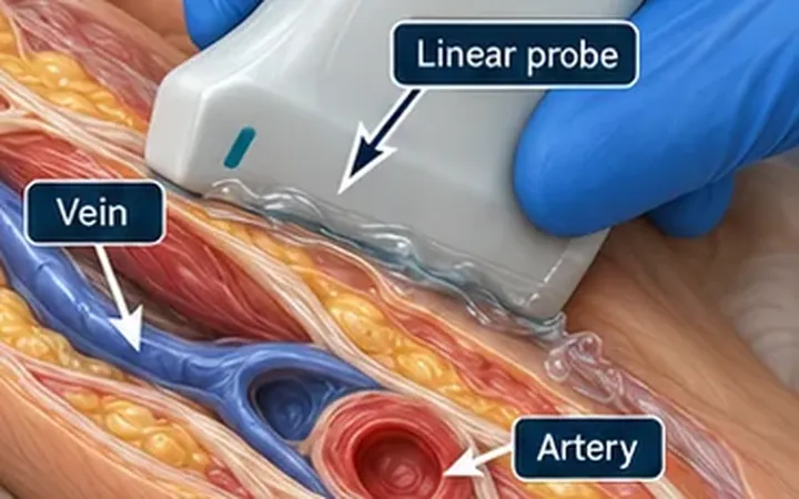
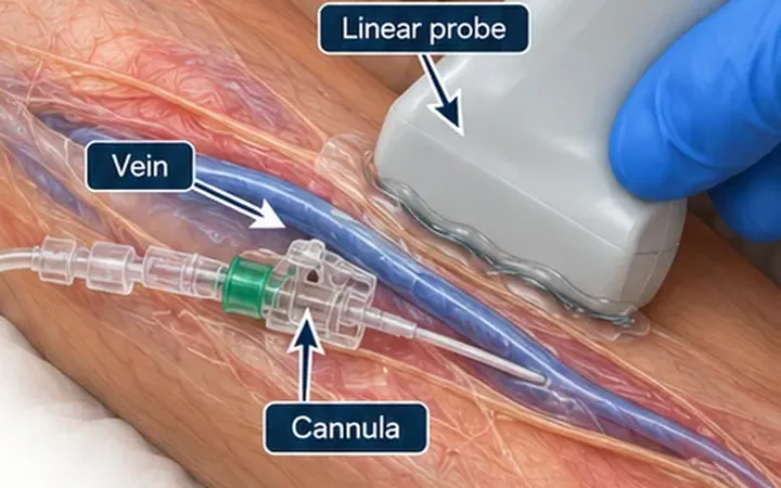
A vein is a thin-walled vessel with low pressure inside it. The probe is a hard object pressed on top. Press even a little too hard and a vein that was easy to see becomes a thin line or disappears. Scanners then conclude the vein is too small, and move on to a worse target.
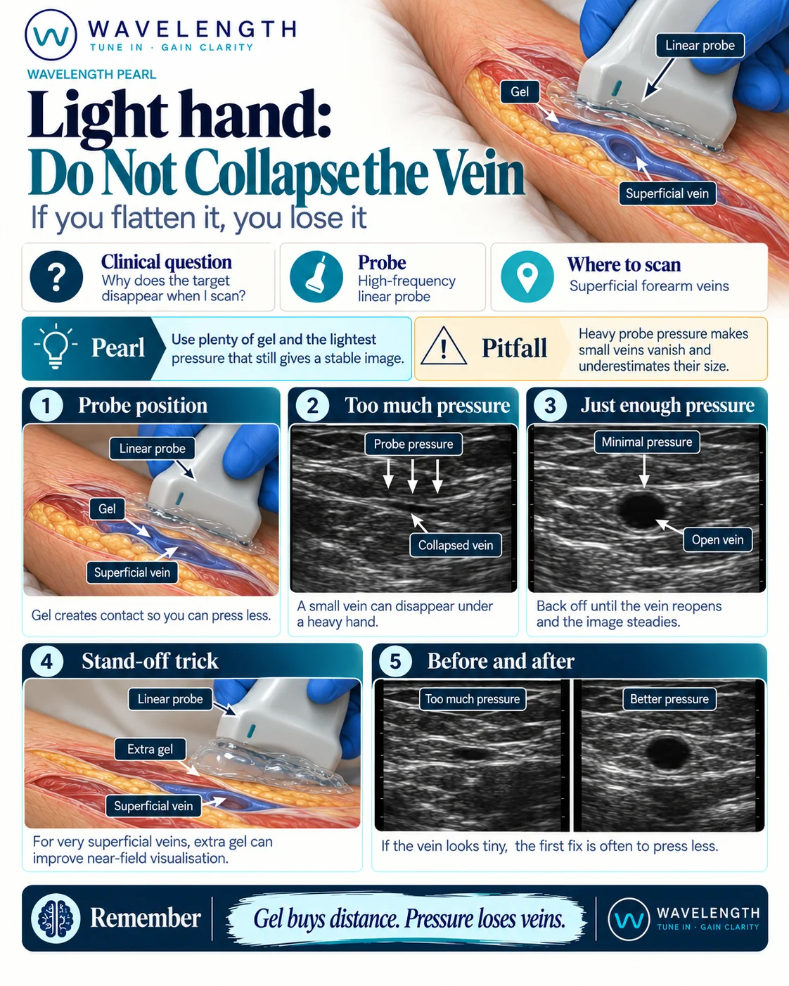
Scan images in the graphic are illustrations, not patient scans.
What to do
- Apply generous gel. Gel carries the sound, so you do not need pressure to make contact.
- Rest the probe on the gel with a light touch.
- If the vein looks tiny or slit-like, lift off until it reopens and the image steadies. The first fix for a small vein is often to press less.
- For a very superficial vein, add a thick layer of gel as a stand-off. The vein moves away from the top of the image, where near-field detail is poor, and into clear view.
- Keep the same light pressure while you slide, and while you puncture.
Reading the view
- Too much pressure. A flat, slit-like vein under the probe, and the artery still round beside it.
- Just enough pressure. A round, open, black vein with the skin line intact above it.
Pressure as a tool
Pressure is useful when you choose it. A vein collapses under gentle pressure and an artery does not. Use that to tell the two apart, then go back to the light touch for the puncture.