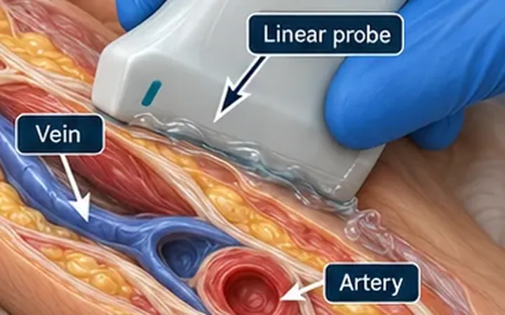
The worst central line complications start with a wire in the wrong place. A wire in the carotid artery, followed by a large dilator and a catheter, can cause a major arterial injury. Flashback, blood colour and a smooth wire pass can all mislead, and a wire can pass through the vein wall into the tissues. Ultrasound shows the wire directly, and it takes a few seconds.
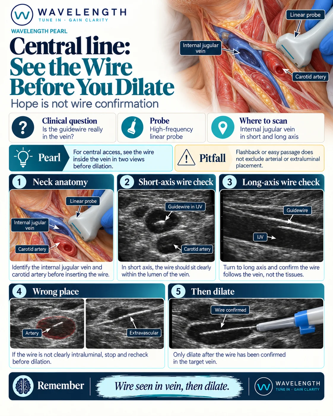
Scan images in the graphic are illustrations, not patient scans.
Before you start
This pearl covers one safety check. It does not teach central venous catheter insertion. Central lines need consent, full sterile precautions, a trained operator or direct supervision, and your trust's policy.
The scan
- Before you prepare the skin, scan the neck. Identify the internal jugular vein and the carotid artery in short axis. The vein is usually lateral to the artery and compresses with gentle pressure.
- Puncture under real-time ultrasound, with the needle tip in view.
- After you pass the wire, put the probe back over the vein in short axis. The wire should sit clearly inside the black lumen of the vein, as a bright dot or short line.
- Turn to long axis and follow the wire along the vein. It should run within the lumen, not in the tissues beside it.
- If you cannot see the wire clearly inside the vein, stop and recheck before you dilate.
- Dilate only once the wire has been seen in the target vein.
Reading the view
- Wire in the vein. Bright echo inside a black, compressible lumen, in two views.
- Wire in the artery. Bright echo inside the pulsating, non-compressible carotid.
- Extravascular wire. A bright line running outside the lumen, in the tissue beside the vein.
What the wire check does not show
A wire inside the internal jugular vein does not tell you where the catheter tip ends up. After insertion, confirm the tip position and look for complications such as pneumothorax, in line with your trust's policy.
The recommendations
The 2019 Society of Hospital Medicine position statement recommends real-time ultrasound for internal jugular vein cannulation, and recommends that you visualise the needle tip and the guidewire in the target vein before dilation. It accepts either a short-axis or a long-axis approach.