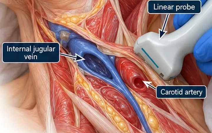
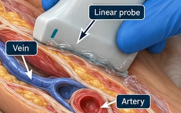
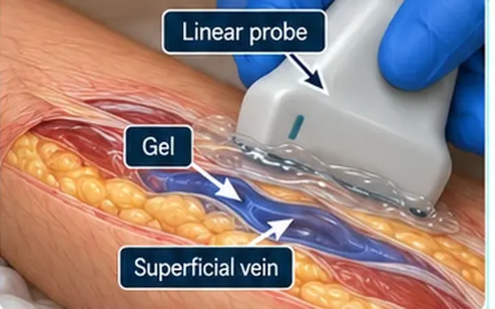
A cannula that gives a flash and flushes easily may still lie outside the vein. A cannula placed in a difficult patient, or in a vein that you found with ultrasound, is the one you most need to trust. Watching a small flush on the screen answers the question in seconds, before you run a drug through it.
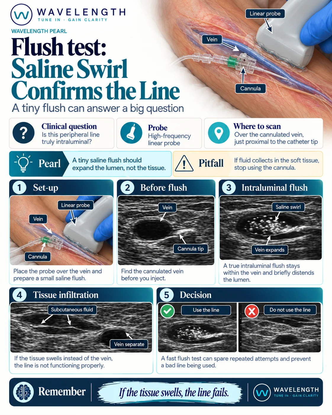
Scan images in the graphic are illustrations, not patient scans.
The scan
- Hold a high-frequency linear probe over the cannulated vein, just proximal to the catheter tip. Find the vein and the cannula tip before you flush.
- Check that you are looking at a vein: round or tubular, compressible and not pulsatile.
- Flush a small volume of saline through the cannula while you watch the vein.
- Look inside the lumen. A swirl of bright echoes, flowing away from the tip, means the fluid is in the vein. The vein may briefly distend.
- If the lumen stays empty and fluid collects in the soft tissue, the cannula is not in the vein. Stop using it.
Reading the view
- Intraluminal flush. A bright swirl of microbubbles in the black lumen, which stays within the vein.
- Tissue infiltration. A dark or mixed pool of fluid in the subcutaneous tissue, with the vein separate and unchanged.
What it does not replace
The flush test adds to the usual checks and does not replace them. Look for blood return, ease of flushing, pain and swelling. Before you infuse an irritant or a vesicant drug, follow your trust's policy and confirm placement with extra care. A swirl in an artery can look similar, so confirm that the vessel is a vein first, by compression.
The evidence for ultrasound confirmation of a flush in adults is limited. Studies exist in specific groups, such as children, using other ultrasound methods. Treat the flush test as a practical bedside check.