Wavelength Pearls
Ultrasound-guided vascular access.
Peripheral cannulation, central line safety and arterial lines with ultrasound.
Ultrasound helps most when a vein is hard to see or feel. These pearls follow a cannulation from the first scan to the last check: centring the vein, using a light hand, filling the vein before you scan, stopping at the near wall, rescuing a catheter that will not thread and confirming the line with a flush.
They also cover two procedures where the image prevents harm: confirming the guidewire is inside the internal jugular vein before you dilate, and choosing a straight stretch of radial artery for an arterial line. Each pearl ends with a short test. Follow your trust's policy on training, supervision and sterile technique.
8 pearls
One technique at a time.
Vascular access
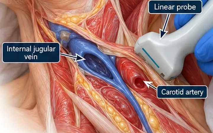
Central line: see the wire before you dilate
Flashback and easy wire passage do not prove the wire is in the vein. See the wire inside the internal jugular vein in two views before you dilate.
Vascular access
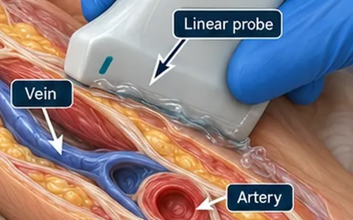
Peripheral access: centre the dot
Before you puncture, bring the vein to the middle of the screen, use the lightest pressure that keeps it round, and prove you can keep it there. A centred vein gives a straighter, safer needle path.
Vascular access
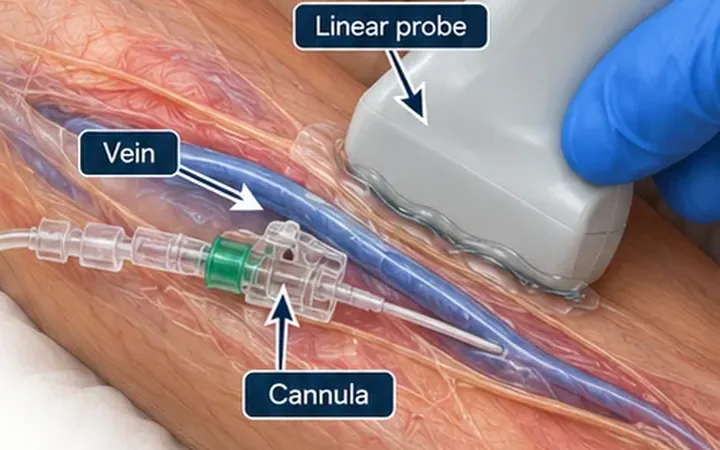
Flush test: saline swirl confirms the line
A small saline flush through the cannula, watched with a linear probe over the vein, tells you whether the fluid fills the lumen or the soft tissue. If the tissue swells, the line fails.
Vascular access
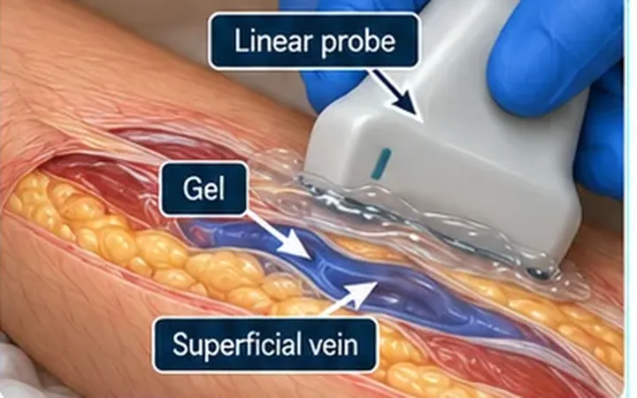
Light hand: do not collapse the vein
Veins hold low pressure, so a heavy probe flattens them and the target seems to vanish. Use plenty of gel and the lightest pressure that still gives a stable image.
Vascular access
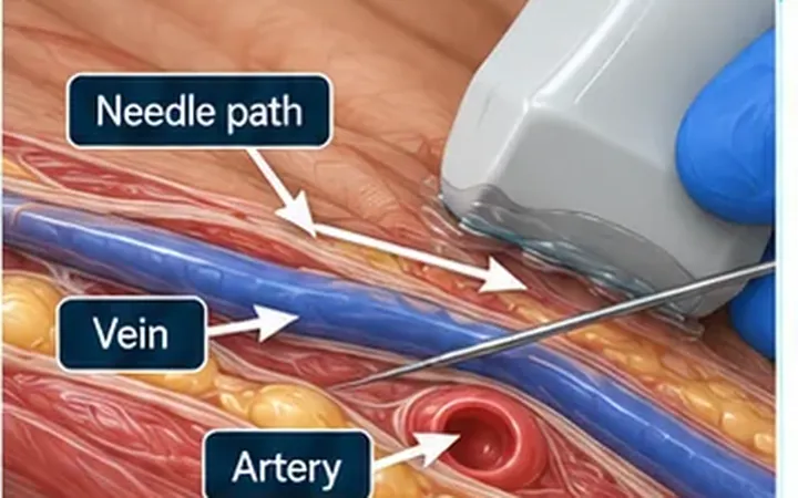
Posterior wall puncture: stop at the near wall
A flash of blood does not mean the needle is in the lumen. Slow down as the tip reaches the near wall, watch it enter the lumen, then stop before the back wall.
Vascular access
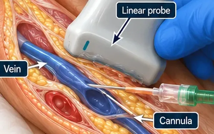
Threading failure: rescue in long axis
Short axis finds the vein and long axis saves the line. When the tip is in the vein but the catheter will not advance, rotate to long axis and look for wall tenting, a valve or a poor angle.
Vascular access
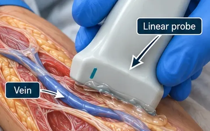
Distension tricks: make the vein come to you
Before you scan, fill the veins. A tourniquet, a dependent arm, warmth and a relaxed squeeze of the hand turn a marginal vein into a usable one.
Vascular access
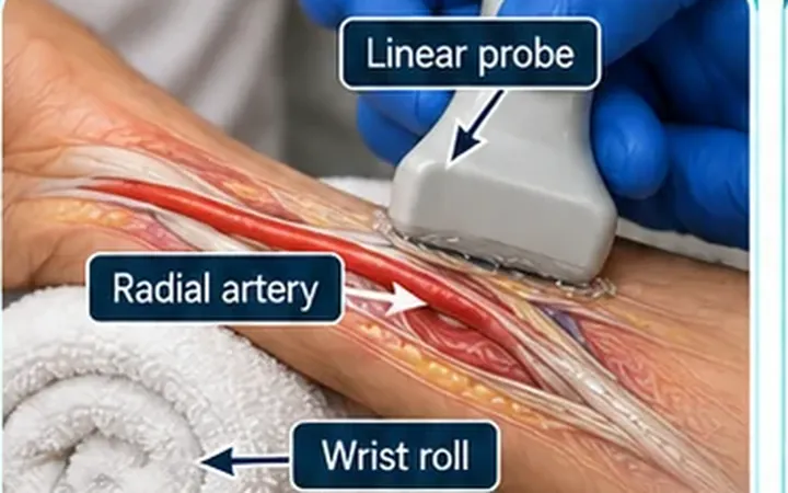
Radial arterial line: trace the pulsation
Pick a straight, superficial stretch of radial artery, steady the wrist and enter at a low angle. If the puncture works but the catheter will not go in, rotate to long axis.
All Wavelength Pearls · Question bank · Log these scans in the free POCUS logbook
Practise it with us
One instructor to every four or five delegates, FAMUS-accredited instructors and RCEM-approved ultrasound supervisors, and long, supervised time on the probe.
Newsletter
Get the next one by email.
One email a week: a practical scanning skill, new Learn posts and course dates before they go public.