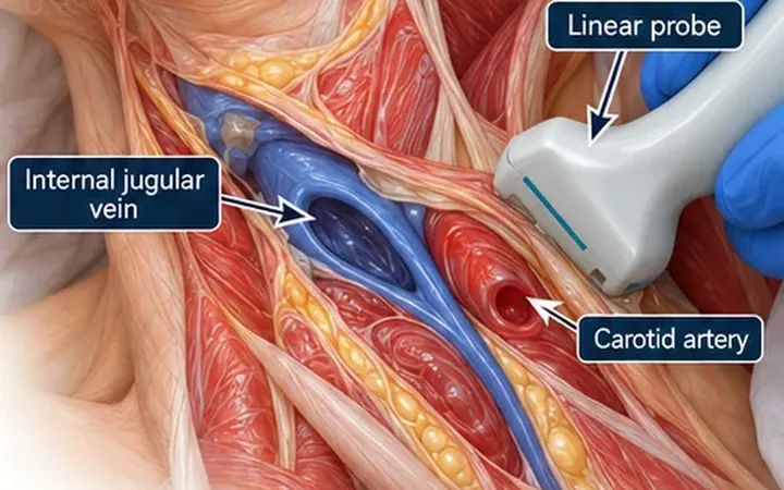
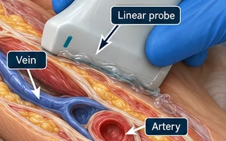
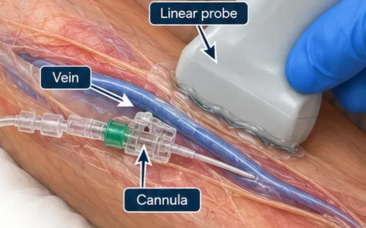
The commonest way to lose a good vein is to go through it. The needle enters the lumen, the operator pushes on, and the tip passes through the back wall. A flash of blood can still appear, because the bevel can sit partly in the lumen and partly outside it. The catheter then fails to thread, or the vein blows and the patient has a bruise.
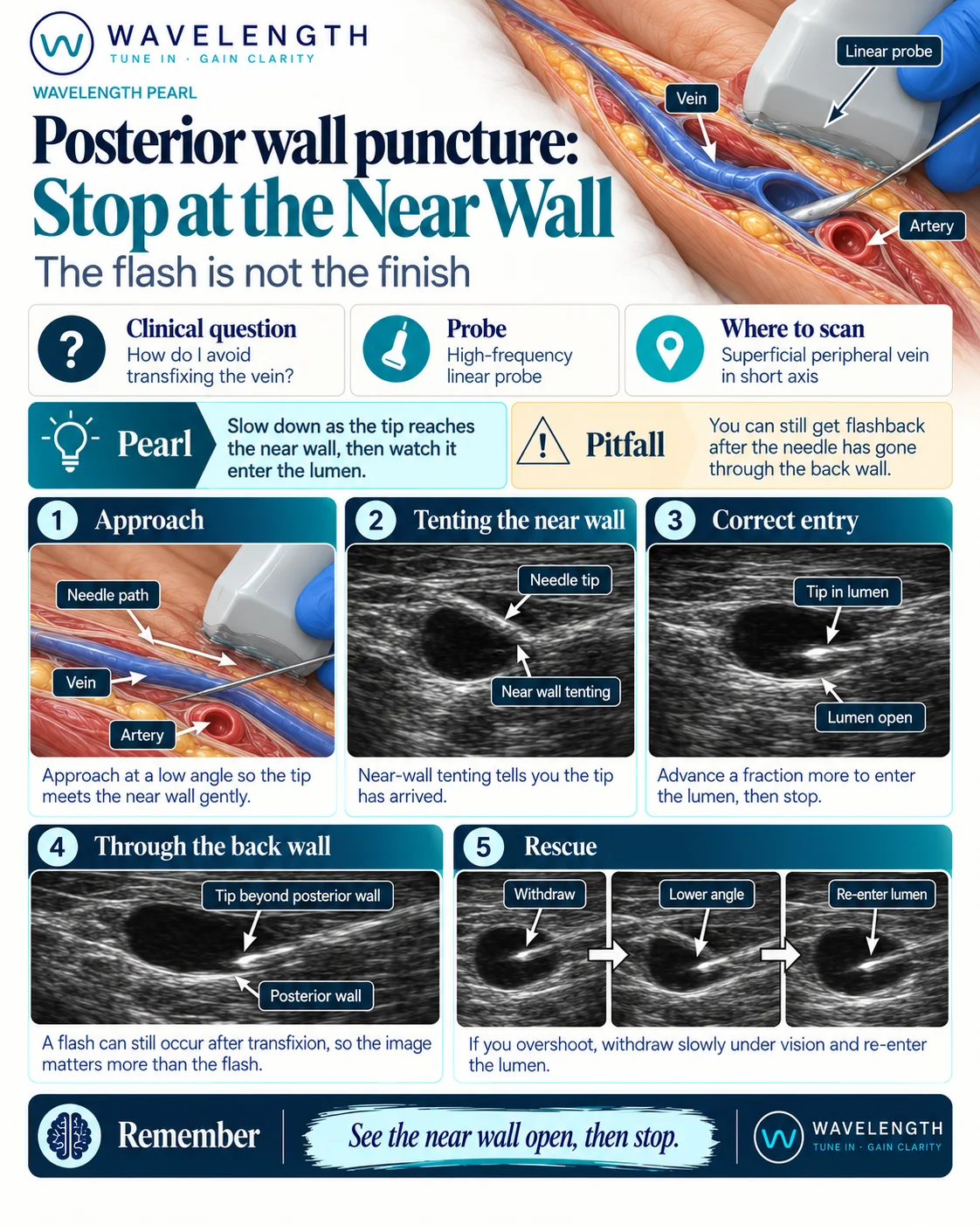
Scan images in the graphic are illustrations, not patient scans.
The scan
- Scan the vein in short axis. Keep it centred and round with light pressure.
- Approach at a low angle, so the tip meets the near wall gently.
- Follow the tip as it advances. Slide the probe a little after the needle so the tip stays in view.
- When the tip reaches the near wall, slow down. The wall tents, which tells you the tip has arrived.
- Advance a fraction more, until the tip is inside the lumen. Then stop.
- Confirm by image, not by flash, before you advance the catheter.
If you overshoot
- Stop and keep the probe still, with the tip in view.
- Withdraw slowly under vision until the tip is back inside the lumen.
- Lower the angle and re-enter.
- Check the tip position again before you advance the catheter.
Reading the view
- Near-wall tenting. The wall bends in toward the lumen under the needle tip. It is a sign the tip is at the wall, not in the vein.
- Tip in lumen. A bright dot inside a black, open lumen.
- Through the back wall. The bright tip lies beyond the posterior wall, even though blood is still returning.
Why it matters at the elbow
In the antecubital fossa and the upper arm, an artery or a nerve often lies beneath the vein. A needle that passes through the back wall can injure it. Take extra care to see the tip and depth in these areas.