The retropharyngeal space lies between the back wall of the pharynx and the prevertebral muscles in front of the spine. In children under five it holds lymph nodes that drain the nose and throat; after a cold or tonsillitis they can suppurate and form an abscess. Adults get it after a penetrating injury such as a fish bone, after instrumentation, or with immunosuppression. It can threaten the airway, spread down into the mediastinum or involve the internal jugular vein.
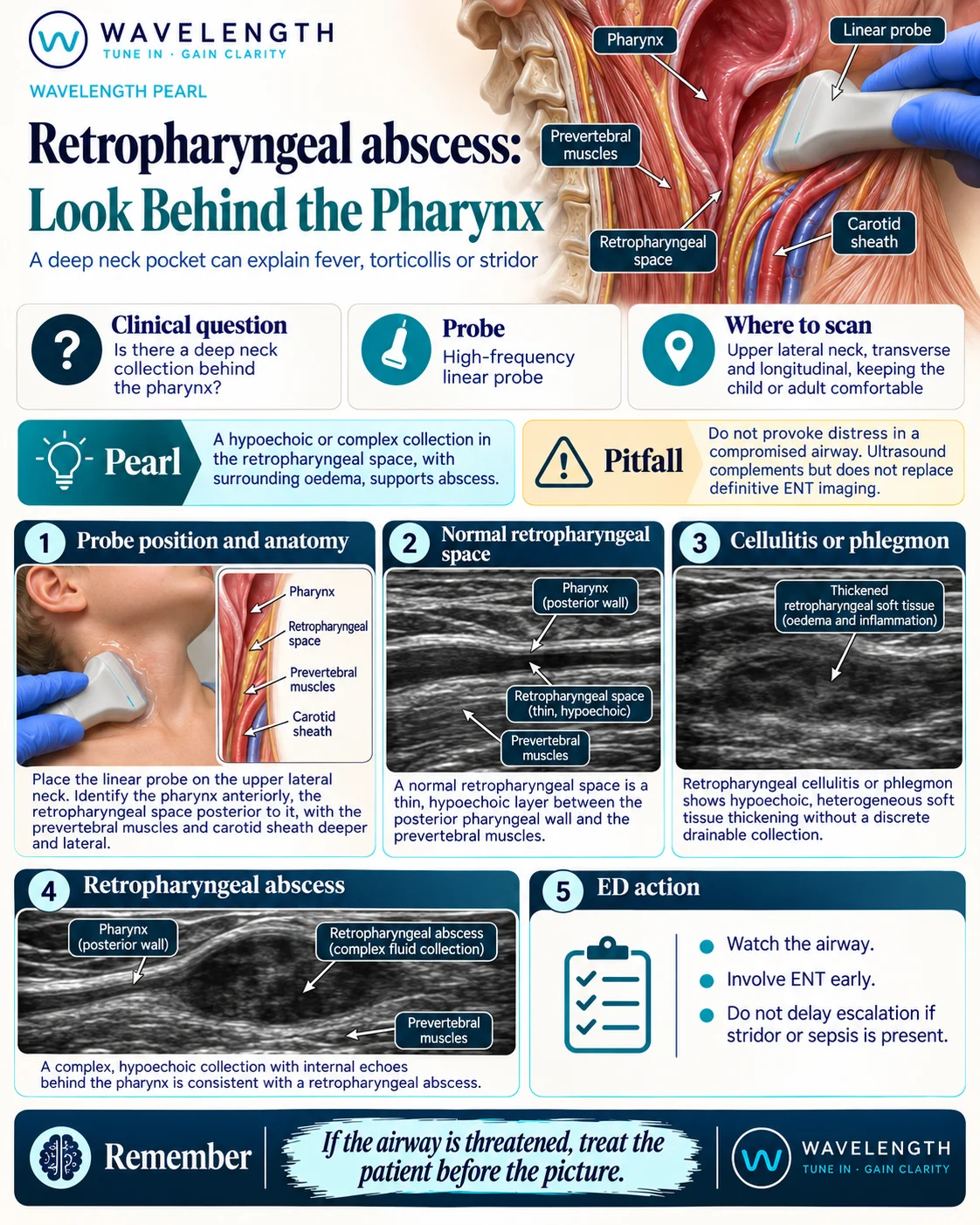
Scan images in the graphic are illustrations, not patient scans.
The scan
- Scan only if the patient is calm and the airway is safe. A child with stridor or drooling stays upright with a parent, and the airway team comes first.
- Use a high-frequency linear probe on the upper lateral neck, in front of the sternocleidomastoid, in transverse and long axis.
- Identify the carotid artery and internal jugular vein with colour Doppler, and the pharynx medially.
- Look medial and deep to the vessels, behind the pharynx and in front of the prevertebral muscles.
- Scan both sides and compare.
Reading the view
- Normal: a thin layer of soft tissue between the back of the pharynx and the prevertebral muscles.
- Cellulitis or phlegmon: thickened, dark, heterogeneous soft tissue with no discrete pocket.
- Abscess: a hypoechoic or complex collection with internal echoes behind the pharynx, often with surrounding oedema and no flow inside it.
- Check the vein: a compressible internal jugular vein with flow; an echogenic, non-compressible vein suggests thrombosis.
Action
Senior airway help and ENT early. Contrast-enhanced CT of the neck defines the collection and its spread when it is safe to transfer. Give intravenous antibiotics. Ultrasound can help ENT plan or guide drainage once the collection is confirmed.