Lung sliding is the first question in a suspected pneumothorax. The answer depends on looking at the right line. Many new scanners place the probe along the ribs, pick up the surface of a rib, see no movement, and call it absent sliding.
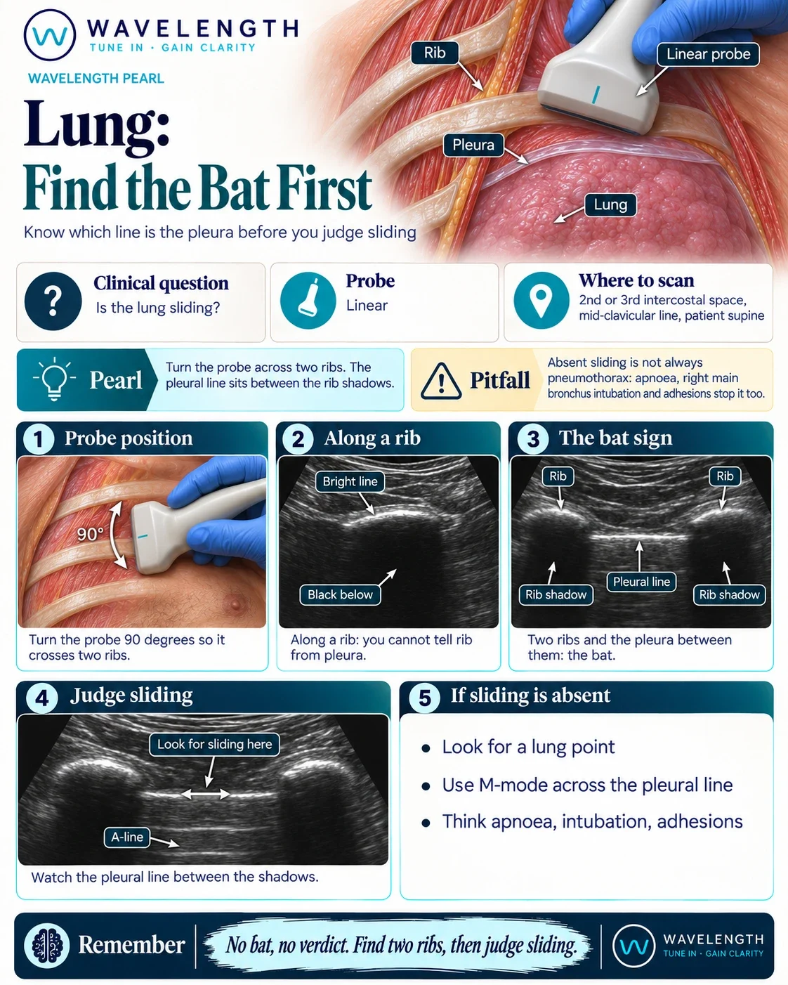
Scan images in the graphic are illustrations, not patient scans.
Before
With the probe lying along a rib, the beam hits bone. You see one bright, curved line with a black shadow filling the screen beneath it. The surface of a rib and the pleura both look like a bright line, so you have no way to tell which one you are watching. A rib never slides.
The move
- Start with the linear probe in the second or third intercostal space, mid-clavicular line, with the patient supine.
- Turn the probe 90° so it lies head to foot and crosses two ribs.
- Point the marker to the head.
- Hold the probe still, with light pressure, and let the patient breathe.
After
Now you see two ribs, each with its own shadow, and a bright horizontal line between them, sitting slightly deeper than the top of the ribs. This is the pleural line. The ribs form the wings and the pleura forms the body of a bat. Watch the pleural line between the shadows. Shimmering movement along it is lung sliding.