Adult epiglottitis presents as a severe sore throat, pain on swallowing and a muffled voice, often with a normal-looking pharynx. Drooling, stridor or sitting forward are signs of a threatened airway. Direct laryngoscopy by ENT is the usual test. In a stable adult, the epiglottis can also be seen with a linear probe on the front of the neck. Small studies have found it thicker in epiglottitis, but they used different cut-offs, so there is no single agreed number.
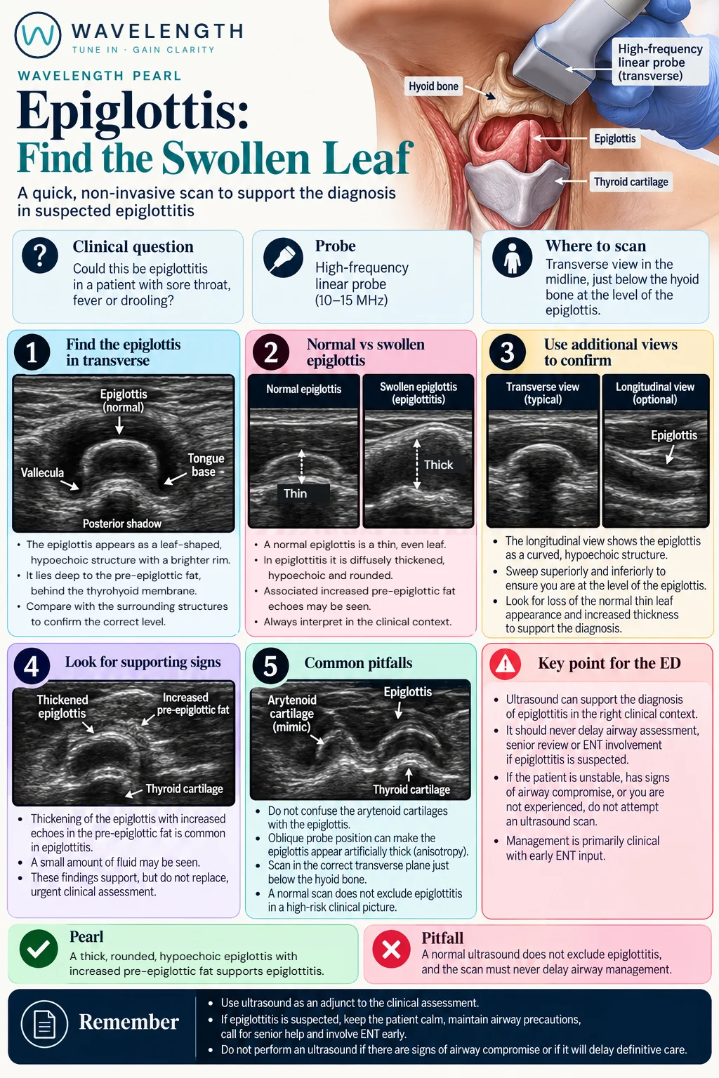
Scan images in the graphic are illustrations, not patient scans.
The scan
- Do not scan a patient with stridor, drooling, distress or any sign of airway compromise. Call for senior, anaesthetic and ENT help at once.
- Do not lie the patient flat or upset them. Let them sit as they are comfortable. Avoid scanning distressed children.
- Use a high-frequency linear probe in transverse, in the midline, between the hyoid bone and the thyroid cartilage.
- Tilt the probe towards the head until a curved, dark band appears deep to the bright pre-epiglottic fat: the epiglottis.
- Sweep up and down to stay at its level, and confirm it in a longitudinal view.
Reading the view
- Normal: a thin, even, curved dark band, like a leaf in cross-section.
- Epiglottitis: a thickened, rounded, darker epiglottis, sometimes with brighter, swollen pre-epiglottic fat in front of it.
- Mimics: the arytenoid cartilages lie lower, and an oblique probe angle makes a normal epiglottis look thick.
Action
Suspected epiglottitis needs senior review, early ENT assessment and an airway plan, with the patient kept calm and upright. Start intravenous antibiotics and steroids according to local guidance. Ultrasound is an adjunct in stable adults only.