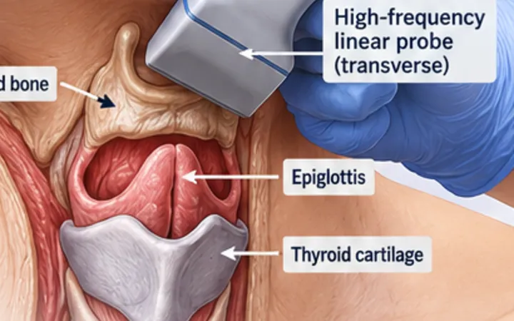
Feeding or flushing through a nasogastric tube that sits in the airway causes serious harm, and in England it is a Never Event. UK guidance confirms position with an aspirate pH of 1 to 5.5, or with a chest X-ray read by a competent person. Ultrasound is not one of the confirmation methods. It is still useful: in the neck it shows whether the tube has gone down the oesophagus, the same anatomy you use when you check an endotracheal tube.
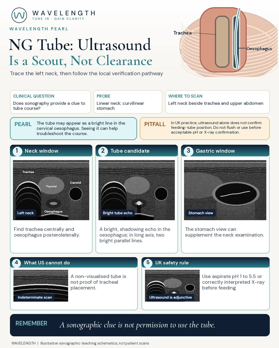
Scan images in the graphic are illustrations, not patient scans.
The scan
- Use a high-frequency linear probe in transverse on the left side of the neck, at the level of the thyroid.
- Find the trachea in the midline: a bright curved line with reverberation below it.
- Find the oesophagus behind and to the left of the trachea: a round structure with a dark muscular wall and a bright centre. A swallow makes it move.
- Look for the tube inside it, and turn to long axis along it.
- Optionally, scan the stomach with a curvilinear probe below the left costal margin.
Reading the view
- Tube in the oesophagus: a bright echo with a shadow inside the oesophagus in transverse, and two bright parallel lines in long axis.
- Stomach: a bright line within fluid in the stomach, if gas does not block the view.
- Not seen: gas, a collapsed oesophagus or a thin tube can hide it.
Action
Follow the local nasogastric tube policy. Do not flush or feed until the aspirate pH is 1 to 5.5, or a chest X-ray read by a competent person confirms the position. Do not use the whoosh test, litmus paper or the patient's comfort to check placement.