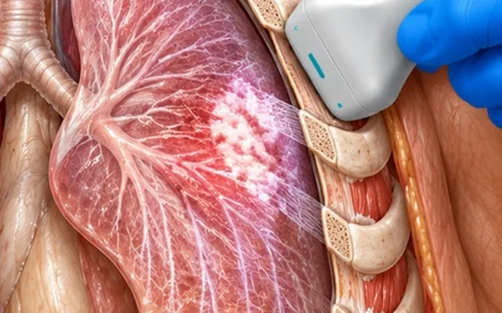
Finding a pleural effusion is the first step, shown in the pleural effusion pearl. The next is its character. Ultrasound shows septations and loculations better than CT, and they change the plan: a septated effusion in a febrile patient is pleural infection until proven otherwise.
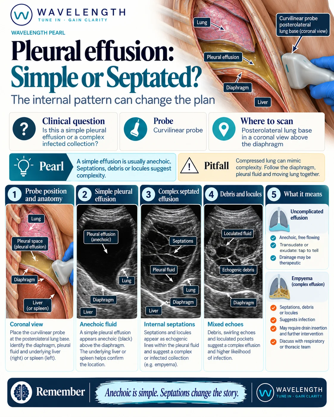
Scan images in the graphic are illustrations, not patient scans.
The scan
- Sit the patient up if you can, or scan the posterolateral chest with the patient supine.
- Use the curvilinear probe in the coronal plane above the diaphragm, with the liver or spleen below.
- Find the diaphragm, the fluid and the lung within it.
- Turn up the gain a little and look for echoes, strands and walls in the fluid.
- Sweep through the whole collection: septations may be in only one part.
Reading the view
- Anechoic: black fluid. Can be a transudate or an exudate, so it does not tell you which.
- Complex non-septated: echoes or swirling particles within the fluid. An exudate, such as a parapneumonic effusion or blood.
- Complex septated: strands or a honeycomb of septations. An exudate, often a complicated parapneumonic effusion or empyema.
- Homogeneously echogenic: uniformly grey fluid, which can be pus or blood.
- Thickened, nodular pleura: raises the question of cancer.
Action
A septated or complex effusion in an unwell patient needs a diagnostic tap under ultrasound guidance and discussion with the respiratory team: a pH below 7.2, pus, or organisms suggest the effusion needs a drain. Use ultrasound to mark or guide every pleural procedure.