Back pain with new bladder symptoms, saddle numbness or leg weakness raises the possibility of cauda equina syndrome. The clinical signs are often soft, and patients find bladder symptoms hard to describe. A bladder scan after voiding gives one number you can measure, repeat and hand over. It supports the decision for urgent MRI. It does not make it.
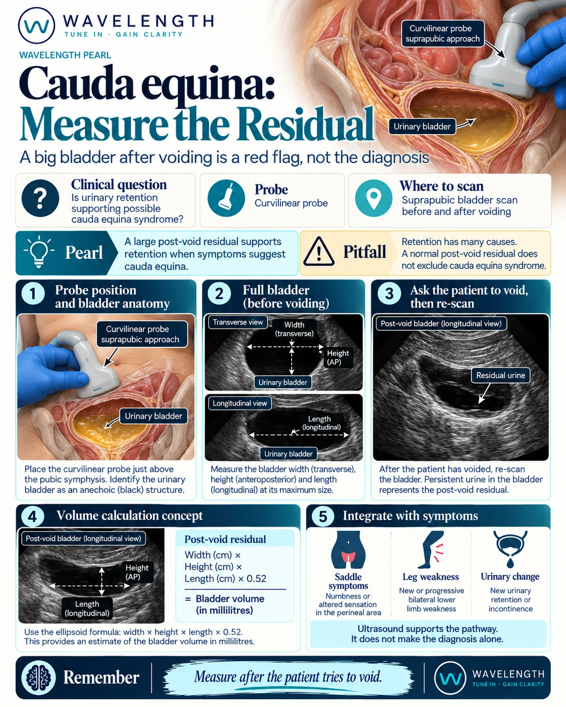
Scan images in the graphic are illustrations, not patient scans.
The scan
- Ask the patient to pass urine first, in private and as normally as they can. Note the time.
- Scan within about 10 minutes of voiding. Use the curvilinear probe just above the pubic symphysis, angled down into the pelvis.
- In transverse, find the widest view of the bladder and measure the width and the front-to-back height.
- Turn the probe to sagittal and measure the longest length.
- Volume in millilitres is about width × height × length (in centimetres) × 0.52. A bladder scanner gives the same estimate automatically.
- If the bladder is very full before voiding, scan it then too. A painless large bladder is itself a warning sign.
Reading the view
- Small residual: the bladder has emptied well. This is reassuring for retention, not for cauda equina syndrome.
- Large residual: a bladder that stays full after a real attempt to void. Many UK pathways treat a residual of more than about 200 mL as significant, but no single threshold rules the syndrome in or out.
- Painless retention: a large residual without the discomfort you would expect is more worrying than a large residual in pain.
- Upstream: with a large bladder, look at both kidneys for hydronephrosis, as in the urinary retention pearl.
Action
Follow your local suspected cauda equina pathway. If the history or examination suggests the syndrome, arrange urgent MRI whatever the residual. Record the residual volume, the time since voiding and how it was measured, so the next clinician can compare.