Plantar heel pain is very common, and most of it is plantar fasciitis, worst with the first steps in the morning. A rupture is rarer: a sudden pop and sharp pain under the foot, sometimes after a steroid injection. A quick comparison of both heels with a linear probe separates them.
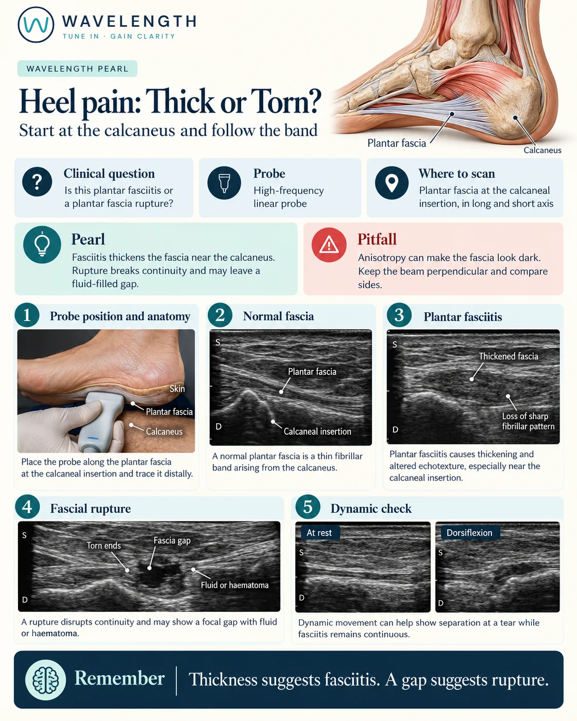
Scan images in the graphic are illustrations, not patient scans.
The scan
- Lay the patient prone with the feet over the end of the bed, or with the foot resting on its side.
- Place a high-frequency linear probe in long axis on the sole, over the heel, so the calcaneus is a bright curve.
- Find the plantar fascia as a bright, fibrillar band arising from the calcaneus and running forwards.
- Measure its thickness where it leaves the bone.
- Follow it forwards, then scan in short axis.
- Compare with the other heel. Gently bend the toes upwards to tension the fascia if you suspect a tear.
Reading the view
- Normal fascia: a thin, bright fibrillar band, usually less than about 4 mm thick at the heel.
- Plantar fasciitis: thicker than about 4 mm, darker, with loss of the fibrillar pattern, usually at the heel.
- Rupture: a break in the fascia, often a few centimetres forward of the heel, with fluid or haematoma in the gap. The gap opens as the toes are bent upwards.
Action
Plantar fasciitis is managed with stretching, footwear advice and time, through your local pathway. A rupture needs offloading in a boot and follow-up with orthopaedics or podiatry. Heel pain with swelling, night pain or systemic symptoms needs other causes considered, such as a calcaneal stress fracture or infection.